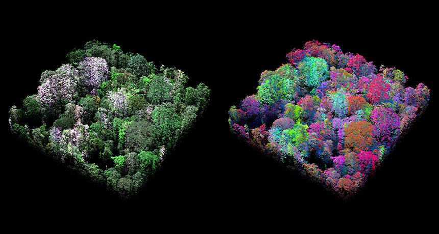
To some forest creatures, a tree is a home. To scientists, it’s a beacon. A new way of mapping forests from the air by measuring chemical signatures of the tree canopy is revealing previously unrecognized biodiversity.
The swath of tropical forest covering the Peruvian Andes Mountains and the Amazon basin is one of the most biodiverse places on Earth. But it’s such a wild and remote region that variation within the forest is hard to spot. “If you look in Google Earth, it just looks like a big green blanket,” says study coauthor Greg Asner, an ecologist at the Carnegie Institution for Science in Stanford, Calif.
Up close, it’s a different story. Each tree species has a distinctive set of chemical traits, such as levels of nutrients like nitrogen and phosphorus in the leaves. Collectively, those characteristics can reveal a lot about the makeup of the forest. To peek beneath the green blanket, Asner and colleagues divided 76 million hectares of forest into 100-kilometer squares. The researchers measured levels of water, nitrogen, phosphorus and calcium in the trees’ leaves via aircraft by measuring the wavelengths of light reflected by the forest canopy, taking samples from small areas of each square. They also mapped leaf levels of lignins and polyphenols, two chemicals used for defense. Using that data, the scientists identified 36 unique types of forest — a much more nuanced view than the broad categories currently used for classification, the researchers report January 27 in Science. They parceled those highly specific forest types into six groups that roughly aligned with the country’s topography and geography. Parsing out these differences in forests at such a fine scale is important for guiding conservation efforts, Asner says. A particular spot might appear at a distance to be the same as its surroundings but may actually contain species found nowhere else.
The team is now carrying out similar studies in northern Borneo and Ecuador. Eventually the researchers hope to boost their sensors into orbit to map biodiversity around the globe.
There are few simple answers in science. Even seemingly straightforward questions, when probed by people in search of proof, lead to more questions. Those questions lead to nuances, layers of complexity and, more often than we might expect, conclusions that contradict initial intuition.
In the 1990s, researchers asking “How do we fight oxygen-hungry cancer cells?” offered an obvious solution: Starve them of oxygen by cutting off their blood supply. But as Laura Beil describes in “Deflating cancer”, oxygen deprivation actually drives cancer to grow and spread. Scientists have responded by seeking new strategies: Block the formation of collagen highways, for instance, or even, as Beil writes, give the cells “more blood, not less.” In “DNA tests inflate species counts,” Tina Hesman Saey reports on the complications of classifying species. Genetic analyses alone, she writes, can detect too many differences, overestimating species numbers. Some tools appear to be, as Darwin would have put it, “hair-splitters” rather than “lumpers.” Identifying species is hard in part because “What is a species?” has no single answer. The notion of reproductive isolation, which splits species according to whether they can produce fertile offspring, has little meaning for asexual organisms, for instance. And isolation itself is a matter of degree. Accounting for speciation in progress is yet another challenge. At what point is a split declared official?
There are countless more examples. The question of what led to the dinosaurs’ demise was solved years ago, we thought. But remaining mysteries inspired a special report earlier this year (SN: 2/4/17, p. 16). And don’t even get me started on “How long does a neutron last?” in Emily Conover’s story “Neutron longevity remains elusive.”
In The Pursuit of Simplicity, physicist Edward Teller described science as a search for simplicity. If that’s the case, the quest is never-ending. With each new insight comes yearning for further insights. I cannot, at this moment, think of a single question that doesn’t demand more exploration. There are answers to be sure, and scientific truths, but for what line of questioning are all the details resolved? Where isn’t there a lingering “why” or “how”? (Think that I’m wrong? Send your ideas to editors@sciencenews.org.)
Wanting to know is innate. Children ask “Why is the sky blue?” or “Where do babies come from?” And parents struggle to answer at the right level of detail. Where does the question begin, and where does it end? What is the best angle of approach? As kids grow up, their questions become more specific, and the answers they receive more complex. Perhaps it’s the students who most appreciate complexity who decide to become scientists. They learn to use the tools of science, which uncovered the complexity in the first place, to try to tame it — diving in ever deeper. And so people end up studying dim and distant galaxies to understand “How did the universe evolve?”, and vats of microbes and methylmercury to ask “How will climate change affect food webs?”
Simplicity may be a gift, but I think complexity is much more interesting. It is one of the great joys of doing science — and of writing about it.
The social lives of macaques and baboons play out in what primatologist Julia Fischer calls “a magnificent opera.” When young Barbary macaques reach about 6 months, they fight nightly with their mothers. Young ones want the “maternal embrace” as they snooze; mothers want precious alone time. Getting pushed away and bitten by dear old mom doesn’t deter young macaques. But they’re on their own when a new brother or sister comes along. In Monkeytalk, Fischer describes how the monkey species she studies have evolved their own forms of intelligence and communication. Connections exist between monkey and human minds, but Fischer regards differences among primate species as particularly compelling. She connects lab studies of monkeys and apes to her observations of wild monkeys while mixing in offbeat personal anecdotes of life in the field.
Fischer catapulted into a career chasing down monkeys in 1993. While still in college, she monitored captive Barbary macaques. That led to fieldwork among wild macaques in Morocco. In macaque communities, females hold central roles because young males move to other groups to mate. Members of closely related, cooperative female clans gain an edge in competing for status with male newcomers. Still, adult males typically outrank females. Fischer describes how the monkeys strategically alternate between attacking and forging alliances.
After forging her own key scientific alliances, Fischer moved on to study baboons in Africa, where she entered the bureaucratic jungle. Obtaining papers for a car in Senegal, for instance, took Fischer several days. She first had to shop for a snazzy outfit to impress male paper-pushers, she says. Fischer and her local guide then shuttled from one government official to another until a well-timed phone call from a local police chief to a key bureaucrat finally produced the forms.
Monkeys get the job done using their own brand of intelligence, Fischer writes. Macaques and baboons navigate their home regions expertly, discern small quantities and object sizes pretty well, and know who’s socially dominant over whom. These abilities are somewhat humanlike, but Fischer draws a bright line between monkeys’ and people’s social lives. Our primate relatives specialize in tracking comrades’ behaviors, she holds, rather than trying to infer others’ plans and desires. And unlike human groups, monkey communities don’t steadily accumulate knowledge and innovations or communicate in languagelike ways, Fischer contends.
So what if monkeys don’t write books or gossip about each other? Their social lives are complex enough to remain largely a mystery to humans, Fischer concludes. The gritty work of conducting long-term studies, especially in the wild, can illuminate the worlds inhabited by monkeys.
Mistakes can be learning opportunities, but the brain needs time for lessons to sink in.
When facing a fast and furious stream of decisions, even the momentary distraction of noting an error can decrease accuracy on the next choice, researchers report in the March 15 Journal of Neuroscience.
“We have a brain region that monitors and says ‘you messed up’ so that we can correct our behavior,” says psychologist George Buzzell, now at the University of Maryland in College Park. But sometimes, that monitoring system can backfire, distracting us from the task at hand and causing us to make another error. “There does seem to be a little bit of time for people, after mistakes, where you’re sort of offline,” says Jason Moser, a psychologist at Michigan State University in East Lansing, who wasn’t part of the study.
To test people’s response to making mistakes, Buzzell and colleagues at George Mason University in Fairfax, Va., monitored 23 participants’ brain activity while they worked through a challenging task. Concentric circles flashed briefly on a screen, and participants had to respond with one hand if the two circles were the same color and the other hand if the circles were subtly different shades.
After making a mistake, participants generally answered the next question correctly if they had a second or so to recover. But when the next challenge came very quickly after an error, as little as 0.2 seconds, accuracy dropped by about 10 percent. Electrical activity recorded from the visual cortex showed that participants paid less attention to the next trial if they had just made a mistake than if they had responded correctly.
The cognitive demand of noting and processing the error seems to divert attention that would otherwise be devoted to the task, Buzzell says.
In real life, people usually have time — even if just a few seconds — to reflect on a mistake before having to make another decision, says Jan Wessel, a psychologist at the University of Iowa in Iowa City. But in some activities such as driving a car or playing a musical instrument, people must rebound from errors quickly while continuing to correctly carry out the rest of the task, he says. Those actions might push the limits of error processing.
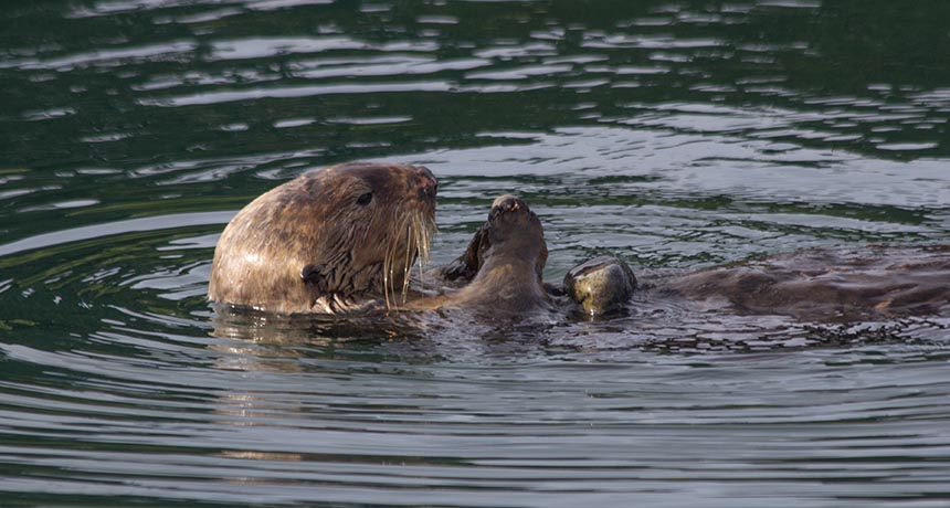
Aside from being adorable, sea otters and Indo-Pacific bottlenose dolphins share an ecological feat: Both species use tools. Otters crack open snails with rocks, and dolphins carry cone-shaped sponges to protect their snouts while scavenging for rock dwelling fish.
Researchers have linked tool use in dolphins to a set of differences in mitochondrial DNA — which passes from mother to offspring — suggesting that tool-use behavior may be inherited. Biologist Katherine Ralls of the Smithsonian Institution in Washington, D.C., and her colleagues looked for a similar pattern in otters off the California coast. The team tracked diet (primarily abalone, crab, mussels, clams, urchins or snails) and tool use in the wild and analyzed DNA from 197 individual otters.
Otters that ate lots of hard-shelled snails — and used tools most frequently — rarely shared a common pattern in mitochondrial DNA, nor were they more closely related to other tool-users than any other otter in the population.
Unlike dolphins, sea otters may all be predisposed to using tools because their ancestors probably lived off mollusks, which required cracking open. However, modern otters only take up tools when their diet requires them, the researchers report March 21 in Biology Letters.
SAN FRANCISCO — When faced with simple math problems, people who get jittery about the subject may rely more heavily on certain brain circuitry than math-savvy people do. The different mental approach could help explain why people with math anxiety struggle on more complicated problems, researchers reported March 25 at the Cognitive Neuroscience Society’s annual meeting.
While in fMRI machines, adults with and without math anxiety evaluated whether simple arithmetic problems, such as 9+2=11, were correct or incorrect. Both groups had similar response times and accuracy on the problems, but brain scans turned up differences.
Specifically, in people who weren’t anxious about math, lower activation of the frontoparietal attention network was linked to better performance. That brain network is involved in working memory and problem solving. Math-anxious people showed no correlation between performance and frontoparietal network activity.
People who used the circuit less were probably getting ahead by automating simple arithmetic, said Hyesang Chang, a cognitive neuroscientist at the University of Chicago. Because math-anxious people showed more variable brain activity overall, Chang speculated that they might instead be using a variety of computationally demanding strategies. This scattershot approach works fine for simple math, she said, but might get maxed out when the math is more challenging.
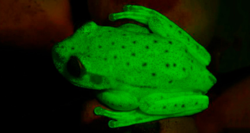
Could fluorescence matter to a frog? Carlos Taboada wondered. They don’t have bedroom black lights, but their glow may still be about the night moves.
Taboada’s question is new to herpetology. No one had shown fluorescence in amphibians, or in any land vertebrate except parrots, until he and colleagues recently tested South American polka dot tree frogs. Under white light, male and female Hypsiboas punctatus frogs have translucent skin speckled with dark dots. But when the researchers spotlighted the frogs with an ultraviolet flashlight, the animals glowed blue-green. The intensity of the glow was “shocking,” says Taboada of the Museo Argentino de Ciencias Naturales “Bernardino Rivadavia” in Buenos Aires. And it is true fluorescence. Compounds in the frogs’ skin and lymph absorb the energy of shorter UV wavelengths and release it in longer wavelengths, the researchers report online March 13 in Proceedings of the National Academy of Sciences. But why bother, without a black bulb? Based on what he knows about a related tree frog’s vision, Taboada suggests that faint nocturnal light is enough to make the frogs more visible to their own kind. When twilight or moonlight reflects from their skin, the fluorescence accounts for 18 to 30 percent of light emanating from the frog, the researchers calculate. Polka dot frogs, common in the Amazon Basin, have plenty to see in the tangled greenery where they breed. Males stake out multilevel territories in vast floating tangles of water hyacinths and other aquatic plants. When a territory holder spots a poaching male, frog grappling and wrestling ensues. Taboada can identify a distinctive short treble bleat “like the cry of a baby,” he says, indicating a frog fight. Males discovering a female give a different call, which Taboada could not be coaxed to imitate over Skype. The polka dot frogs’ courtship is “complex and beautiful,” he says. For instance, a male has two kinds of secretion glands on the head and throat. During an embrace, he nudges and presses his alluring throat close to a female’s nose. If she breaks off the encounter, he goes back to clambering in rough figure eights among his hyacinths, patrolling for perhaps the blue-green ghost of another chance.
A stellar game of chicken between two young stars about 500 years ago has produced some fantastic celestial fireworks, new images released on April 7 by the European Southern Observatory reveal.
Whether or not the stellar duo collided is unclear. But their close encounter sent hundreds of streamers of gas, dust and other young stars shooting into space like an exploding firecracker. Using the Atacama Large Millimeter/submillimeter Array in Chile, John Bally of the University of Colorado Boulder and colleagues made the first measurements of the velocities of carbon monoxide gas in the streamers. From the data, they identified the spot where the stars probably interacted and determined that the encounter ripped apart the stellar nursery in which the stars were born. Such a cataclysmic event flung nursery debris into space at speeds faster than 540,000 kilometers an hour.
The dueling stars were born in a stellar nursery called Orion Molecular Core 1, about 1,500 light-years from Earth behind the Orion Nebula. There, gas weighing 100 suns collapses under its own gravity, making the material dense enough for embryotic stars to take shape. Gravity can pull those stellar seeds toward each other, with some grazing or colliding with each other and violently erupting. In this case, the encounter produced a kick as powerful as the energy the sun emits over 10 million years.
This explosion may have initially released a burst of infrared light lasting years to decades. If so, such spars among young stars might explain mysterious infrared flashes observed in other galaxies, the scientists suggest.
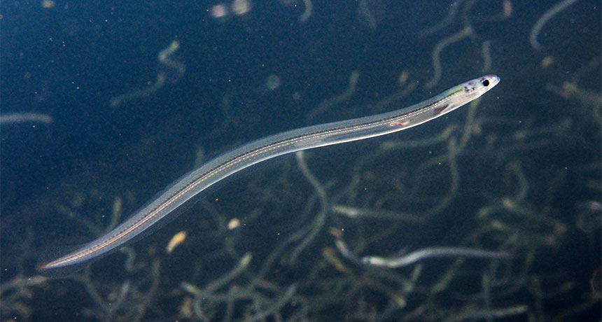
Earth’s magnetic field helps eels go with the flow.
The Gulf Stream fast-tracks young European eels from their birthplace in the Sargasso Sea to the European rivers where they grow up. Eels can sense changes in Earth’s magnetic field to find those highways in a featureless expanse of ocean — even if it means swimming away from their ultimate destination at first, researchers report in the April 13 Current Biology.
European eels (Anguilla anguilla) mate and lay eggs in the salty waters of the Sargasso Sea, a seaweed-rich region in the North Atlantic Ocean. But the fish spend most of their adult lives living in freshwater rivers and estuaries in Europe and North Africa. Exactly how eels make their journey from seawater to freshwater has baffled scientists for more than a century, says Nathan Putman, a biologist with the National Oceanic and Atmospheric Administration in Miami.
The critters are hard to track. “They’re elusive,” says study coauthor Lewis Naisbett-Jones, a biologist now at the University of North Carolina at Chapel Hill. “They migrate at night and at depth. The only reason we know they spawn in the Sargasso Sea is because that’s where the smallest larvae have been collected.”
Some other marine animals, like sea turtles and salmon, tune in to subtle changes in Earth’s magnetic field to help them migrate long distances. To test whether eels might have the same ability, Putman and his colleagues placed young European eels in a 3,000-liter tank of saltwater surrounded by copper wires. Running electric current through the wires simulated the magnetic field experienced at different places on Earth. With no electric current, the eels didn’t swim in any particular direction. But when the magnetic field matched what eels would experience in the Sargasso Sea, the fish mostly swam to the southwest corner of their tank. That suggests the eels might use the magnetic field as a guide to help them move in a specific direction to leave their spawning grounds.
Swimming southwest from the Sargasso Sea seems counterintuitive for an eel trying to ultimately go northeast, Putman says. But computer simulations revealed that that particular bearing would push eels into the Gulf Stream, whisking them off to Europe. Catching a more circuitous ride on a current is probably more efficient for the eels than swimming directly across the North Atlantic, says Putman.
Magnetic fields could help eels stay the course, too. A magnetic field corresponding to a spot in the North Atlantic further along the eels’ route to Europe sent the eels in the tank heading northeast. That’s the direction they’d need to go to keep following the Gulf Stream to Europe.
The researchers did see a fair amount of variation in how strongly individual eels responded to magnetic fields. But that makes sense, says Julian Dodson, a biologist at Laval University in Quebec City who wasn’t part of the study. The Gulf Stream is such a powerful current that the eels could wriggle in a spread of directions to get swept up in its flow.
Now, the researchers are looking at whether adult eels use a similar magnetic map to get back to the Sargasso Sea. Adults follow a meandering return route that might take more than a year to complete, previous research suggests (SN Online: 10/5/16). But whether there’s some underlying force that guides them remains to be seen.
Lab coats aren’t typical garb for mass demonstrations, but they may be on full display April 22. That’s when thousands of scientists, science advocates and science-friendly citizens are expected to flood the streets in the March for Science. Billed by organizers as both a celebration of science and part of a movement to defend science’s vital role in society, the event will include rallies and demonstrations in Washington, D.C., and more than 400 other cities around the world.
“Unprecedented,” says sociologist Kelly Moore, an expert on the intersection of science and politics at Loyola University Chicago. “This is the first time in American history where scientists have taken to the streets to collectively protest the government’s misuse and rejection of scientific expertise.”
Some scientists have expressed concern that marching coats science in a partisan sheen; others say that cat is long out of the bag. Keeping science nonpartisan is a laudable goal, but scientists are human beings who work and live in societies — and have opinions as scientists and citizens when it comes to the use, or perceived misuse, of science.
Typically when scientists get involved with a political issue, it’s as an expert sharing knowledge that can aid in creating informed policy. There are standard venues for this: Professional societies review evidence and make statements about a particular issue, researchers publish findings or consensus statements in reports or journals, and sometimes scientists testify before Congress.
In extreme circumstances, though, scientists have embraced other forms of activism. To broadly categorize, there are:
Celebrity voices In 1938, amid the rise of fascism and use of false scientific claims to support the racism embedded in Nazism, prominent German-American anthropologist Franz Boas released his “Scientists Manifesto.” Signed by nearly 1,300 scientists, including three Nobel laureates, the manifesto denounced the unscientific tenets of Nazism and condemned fascist attacks on scientific freedom. Fear of war of a different sort prompted Albert Einstein, Bertrand Russell and nine other scientists to compose a manifesto in 1955 calling for nuclear disarmament. The Russell-Einstein Manifesto led to the first Pugwash Conference on Science and World Affairs, which sought “a world free of nuclear weapons and other weapons of mass destruction.” Wildlife biologist Rachel Carson eloquently synthesized research on the effects of pesticides in her wildly popular book Silent Spring, published in 1962 (she would later testify before Congress). Despite attacks from industry and some in government, Carson’s work helped launch the modern environmental movement, paving the way for the establishment of the Environmental Protection Agency.
Advocacy groups In the 1930s, chapters of the American Association of Scientific Workers (based loosely on a similar British organization) formed in various cities including Philadelphia, Boston and Chicago. Despite broad goals — promoting science for the benefit of society, stressing public science education, taking a moral stand against government and industry misuse of science — infighting and members’ opposing views limited the group’s effectiveness.
In the decades since, other broadly focused groups — for example, Science for the People (born out of a group started in 1969 by physicists frustrated by their professional society’s lack of action against the Vietnam War), the Union of Concerned Scientists, the American Association for the Advancement of Science — have picked up the banner, speaking out, circulating petitions and more. Single-issue groups such as the Environmental Defense Fund and the Council for Responsible Genetics have proliferated as well.
Protest marchers Many scientists have traded pocket protectors for placards, hitting the streets as concerned scientist-citizens. Academic scientists frequently joined university students in rallies against the Vietnam War in the 1960s and early ’70s. Linus Pauling famously protested nuclear testing in a march outside the White House in 1962 (he was in town for a dinner with the Kennedys honoring Nobel laureates). Carl Sagan was one of hundreds arrested for protesting nuclear testing at a Nevada site in 1987. And plenty of scientist-citizens joined the inaugural Women’s March on Washington in January and the annual People’s Climate March (the 2017 one is scheduled for April 29, just a week after the March for Science).
But the March for Science feels different, say the science historians. Transforming concern into sign-toting, pavement-pounding, slogan-shouting activism is motivated by a collective — and growing — sense of outrage that the federal government is undermining, ignoring, even discarding and stifling science. That’s hitting many scientists not just in their livelihoods, but in the very fabric of their DNA. “Part of [President] Trump’s message is that science is not going to be thought of as part of a collective good that’s essential for decision making in a democracy,” Moore says. “We have not seen this outright rejection of science by the state.”
That rejection has come in many forms, says David Kaiser, a science historian at MIT. “It’s a cluster of issues: cutbacks in basic research across many domains, the censure and censorship regarding data collected by the government or the ability of government scientists to speak, and a range of threats to academic freedom and the research process generally.”
It’s a sign of the times, too, says Al Teich, a science policy expert at George Washington University in Washington, D.C. President Reagan, for example, slashed science in his budget in 1981. But many more people today are aware of science’s role in society, says Teich, the former director for science and policy programs at AAAS. This awareness may be fueling the upcoming march. “The number of people engaged and the range of scientists involved is not something that I’ve ever seen before.”
Measuring the impact of any of these efforts is difficult. They aren’t controlled laboratory experiments, after all. But one thing this march may do is spawn a new form of activism, says Moore: more scientists running for political office.