Human-like robot industry embraces golden age, to feature products with emotions in 5-10 years
NVIDIA CEO Jensen Huang made a splash again in the field of humanoid robots as he was reported to have predicted, in a recent interview, that humanoid robots will soon become mainstream products at prices reaching $10,000 to $20,000 per unit, making them accessible to a wide range of consumers. That will also revolutionize various other industries.
The statement came about a month after he introduced nine humanoid robots at NVIDIA's 2024 GPU Technology Conference (GTC) on March 18. "Building foundation models for general humanoid robots is one of the most exciting problems to solve in AI today," Hung said at that conference.
Hung's judgments, to some extent, can be viewed as a footnote to the golden age of the development of humanoid robots under the stimulus of the boosting of the artificial intelligent (AI) industry in recent years.
Research unveiled during the inaugural China Humanoid Robot Industry Conference in Beijing from April 9 to 10 predicted that the value of the Chinese humanoid robot market will surpass 10 billion yuan, reaching 10.47 billion yuan ($1.45 billion) by 2026, and is anticipated to soar to 119 billion yuan by 2030.
The booming market is seemingly telling people that intelligent humanoid robots that can simulate human thinking and consciousness, as depicted in films like Ex Machina and A.I. Artificial Intelligence. Artificial Intelligence, are almost a reality. But will they come as soon and as easily as predicted by the industry?
Golden age
Humanoid robots are dubbed "humanoid" because they are designed to emulate and potentially surpass human capabilities in form, function, behavior, and even cognitive processes, Zhang Rui, founder and executive director of the Beijing Ironman Technology, told the Global Times.
"Without the need for massive changes to the existing environment, humanoid robots can seamlessly integrate into various scenarios, using their flexible and dynamic execution capabilities to meet complex and dynamic task requirements. Furthermore, their human-like characteristics enable them to easily manipulate human tools, further expanding their application areas," Zhang said.
Therefore, humanoid robots are not only a symbol of technological progress but also a significant force driving future social development, he noted.
Humanoid robots have already been widely applied in various industries, with the aerospace sector being one of the most prominent, according to Zhang. Several countries including the US, Russia, and China have been deeply researching the application of humanoid robots in the aerospace field. These robots are mainly used to replace humans in performing dangerous and complex operations, ensuring the safety of astronauts and improving the efficiency and success rate of space missions.
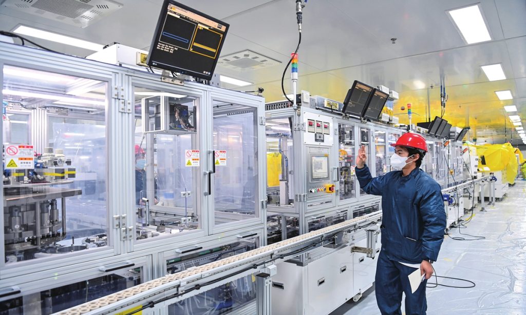
Other major application areas of humanoid robots include border defense and lights-out factories, or smart factories, according to experts.
"The continuous innovation and breakthroughs in AI technology in recent years have indeed provided humanoid robots with more powerful perception, decision-making, and execution capabilities. This allows humanoid robots to more accurately understand human language, recognize environmental information, and make more reasonable decisions and actions," Zhang said.
In the future, Zhang expects humanoid robots to have enormous potential in areas such as general hardware execution, dynamic adaptation, and environmental integration.
The ultimate dream of global humanoid robot developers is to let humanoid robots be integrated into people's daily lives and serve every family, according to Zhang.
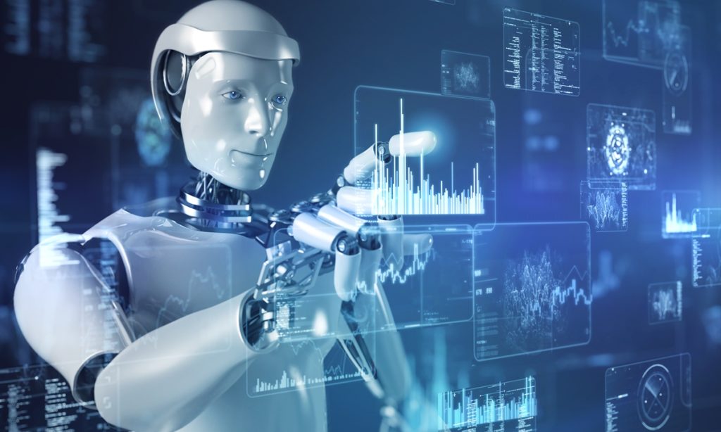
The rapid development of AI technology has played a crucial role in the advancement of humanoid robots. In return, the advancement of humanoid robots is viewed by many industry observers as a major milestone in the AI era, pushing the boundaries of AI research.
Chinese experts and industry observers consider humanoid robots a breakthrough for the "AI Plus" initiative aimed at fostering innovative development in the digital economy. The humanoid robot industry is seemingly embracing its golden age.
Booming market
The thriving progress of humanoid robots is drawing increasing attention from international tech giants. Besides NVIDIA, Tesla is also actively working on a humanoid robot named Optimus. OpenAI, Microsoft, and Amazon founder Jeff Bezos have made substantial investments in humanoid robot startup Figure AI. Additionally, Agility Robotics, backed by Amazon, has established the world's first large-scale humanoid robot production factory in Oregon, the US, capable of producing 10,000 two-legged robots annually.
A group of innovative and competitive Chinese companies have also emerged in the field of humanoid robots with increasing policy emphasis and investment in robot technology in China.
In March, the Beijing humanoid robot innovation center announced that it would soon release the first generation of a universal open humanoid robot body, drawing global attention to the development of China's humanoid robot industry and its supporting industry chain.
Established in November 2023, the center is a major move by Beijing to seize the valuable opportunity to develop the humanoid robot industry.
As the country's first humanoid robot innovation center, the center focuses on the most common five technical tasks for the development of the humanoid robot industry, namely the prototype, general large model, motion control system, tool chain, and operating system. Among them, the prototype is viewed as the body of the humanoid robot, the motion control system is the cerebellum that controls limb movement, and the general large model is the brain.
The Global Times learned from Zhu Songchun, director of the Beijing Institute for General Artificial Intelligence, that the institute would unveil its intelligent humanoid robot prototype TongTong on April 27 at the 2024 Zhongguancun Forum in Beijing which will be held from April 25 to 29. Zhu is a member of the expert committee of the above-mentioned humanoid robot innovation center of Beijing.
According to Zhu, TongTong will be able to complete tasks like assisting humans with pouring tea and water, and offering warm companionship in households, as well as being deployed in multiple tailored scenarios such as nursing homes. In the future, a family for TongTong will be created, including grandparents, younger siblings, and friends.
Among the nine robots showcased at the NVIDIA 2024 GTC, two were developed by Chinese companies, namely H1 from Hangzhou Unitree Robotics and PX5 from Xiaopeng Pengxing.
The Global Times learned from Unitree Robotics that H1 is a full-size humanoid robot capable of running, and equipped with 360 panoramic depth perception. Currently, it can reach a speed of 3.3 meters per second, setting a world record for full-size electric humanoid robots, with a potential speed of up to 5 meters per second.
This robot boasts highly advanced full-body dynamic coordination capabilities, enabling it to dance in groups and execute backflips. As a result, NVIDIA opted to partner with Unitree to collectively propel the global advancement of AI robots.
According to the company's response sent to the Global Times, NVIDIA, a front-runner in GPU and AI chip technology, furnishes Unitree's robots with robust computing capabilities and comprehensive support in deep learning technology.
According to a report by Cailian Press on April 2, the price of H1 is about 650,000 yuan each and 100 units of this model have already been sold.
KUAVO, a product by Chinese company Leju Robot, was sold at about 600,000 yuan, with 30 having been sold. KUAVO is mainly designed for education thus most of its purchasers are colleges and scientific research institutes.
Another Chinese company Kepler Exploration Robotics' product K1 also reportedly sold at a similar price of $20,000, the price of a second-generation Tesla Optimus. The product is designated for smart factories and is expected to be put into mass production in the second half of this year.
Lowering prices will be a major goal for most manufacturers in order to gain market share, Cailian Press reported citing a staff member from Leju.
The company's expansion will be on household service scenarios, with plans to reduce the price of KUAVO to around 200,000 yuan to meet the consumption ability of household users.
Challenges ahead
Although the combination of AI and robots brings many advantages, we must also face the challenges and problems that come with it. First, as the autonomy of robots increases, ensuring the safety and controllability of their behavior has become an urgent issue that needs to be addressed. Second, the integration of AI and robots may lead to the disappearance of a large number of traditional jobs, triggering adjustments in the social employment structure. Additionally, data privacy and security issues are also worth paying attention to, as protecting personal privacy and preventing data leaks have become important topics, Zhang Rui noted.
Zhang believes hardware challenges are a crucial obstacle. While we can achieve various complex functions and performance at the algorithm level, it is often difficult to achieve the desired output power and efficiency in actual robot hardware, he said.
On the other hand, the current progress of AI technology is mainly limited to deepening and innovating at the logical level, with insufficient breakthroughs in thinking and emotional aspects. While the form of robots is malleable, the "spirit" of their internal thought processes and emotions is still an unexplored frontier.
Zhang believes it will take another 5-10 years to achieve a 70-percent similarity with human emotions.
The deep integration of AI and humanoid robots also faces significant challenges. Some experts believe that with the continuous advancement of chips and algorithms, artificial general intelligence (AGI) - a type of artificial intelligence (AI) that can perform as well or better than humans on a wide range of cognitive tasks - will be realized in the near future.
However, Liu Wei, director of the human-machine interaction and cognitive engineering laboratory at the Beijing University of Posts and Telecommunications, pointed out that AGI may be a false proposition. This is not because current AI systems have not reached the level of general intelligence, but because AI fundamentally perform and learns like humans.
Completing the industry chain is also a vital field that Chinese humanoid robot manufacturers and authorities are working on to promote the industry at this rare development time.
In Yizhuang, Beijing, where the Beijing humanoid robot innovation center is located, 110 robotic companies have gathered here, forming a complete industrial chain system covering core components, complete machines, and applications. The whole robot industry in this area is expected to reach a scale of 10 billion yuan in 2025.
With this progress in technology and the gradual improvement of regulations, industry insiders firmly believe that humanoid robots will demonstrate their unique charm in more fields, contributing more to the development of human society.