The Chinese Embassy in the Philippines released details about the relevant communication between China and the Philippines in terms of managing the situation at Ren'ai Jiao. The facts are clear and backed by hard evidence that cannot be denied, Chinese Foreign Ministry spokesperson Lin Jian told a press conference on Wednesday, urging the Philippines to honor its commitment, and stop maritime infringement and provocation at once.
On Tuesday, Bloomberg cited a Chinese official on background, said a transcript of the supposed recording of a phone call with Western Command Commander Vice Admiral Alberto Carlos "may be released to the public within days." In the transcript, Carlos agreed to a "new model" for resupply missions concerning Ren'ai Jiao.
The transcript which documented the phone conversation between the Chinese side and Carlos on January 3 has been confirmed to be true, the Global Times has learned from a source familiar with the affair on Wednesday. Following the conversation, the Philippine side adhered to the "new mode" in the subsequent resupply mission, only delivering essential daily supplies to the grounded warship, and notified the Chinese side in advance.
Based on the "new model" arrangement and humanitarian principle, the Chinese side permitted the Philippine resupply operation, said the source.
However, thereafter, the Philippine side reneged on its promise. Not only did it fail to notify the Chinese side in advance of its resupply activities, but it also attempted to transport construction materials to the illegally grounded vessel, deliberately causing trouble and maliciously hyping up the situation.
The China Coast Guard has firmly restricted the Philippine's illegal resupply activities, according to the source.
Foreign Ministry spokesperson Lin Jian said at the Wednesday press conference that the Philippine side has insisted on denying these objective facts and seeks to mislead the international community. This hurts its own credibility and puts peace and stability in the South China Sea in jeopardy.
China urges the Philippines to honor its commitment, stop maritime infringement and provocation at once, and return to the right track of properly handling disputes with China through dialogue and consultation, Lin noted.
The Philippines' recent denials of the "new model" and the "gentlemen's agreement" reflect the chaos and management confusion as well as multiple conflicting stances within the Philippine government regarding its policies toward China, Ge Hongliang, deputy director of the College of ASEAN Studies at Guangxi University for Nationalities, told the Global Times.
Manila's repeated violations of the gentlemen's agreement between the two sides, or in other words, consensus reached through consultations over dispute management, weaken its claim of seeking a peaceful resolution to the dispute. Its accusing China of "jeopardizing regional peace and security" doesn't hold water, Ge said.
Unlike the Philippines, which is becoming a pawn of the US in the latter's competition with China in the region, China does not wish to see further deterioration of the situation in the South China Sea. China will continue to promote the peaceful resolution of South China Sea disputes through bilateral channels, Ge said.
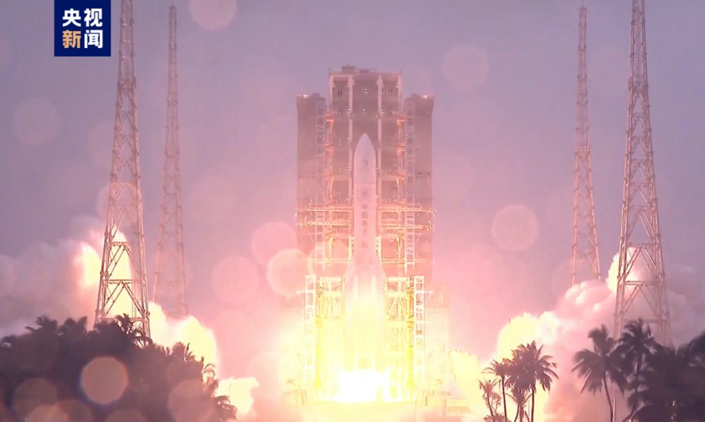
China has made another historic stride in its deep space endeavors on Friday, as the Long March-5 Y8 carrier rocket blasted off at 5:27 pm from the Wenchang Space Launch Site in South China’s tropical island of Hainan, sending the Chang’e-6 lunar probe onto its odyssey in the world’s first ever attempt to bring back lunar samples from the far side of the Moon.
The China National Space Administration (CNSA) confirmed the success of the launch after two pairs of solar panels of the spacecraft opened smoothly.
The round trip of Chang'e-6 to the moon and back will take about 53 days, more than double the duration of its predecessor Chang’e-5, which returned samples from the near side of the moon in some 23 days, media reported.
The longer duration also indicates more complex flight stages – researchers have designed 11 stages for Chang’e-6, including launch and orbit insertion, lunar transfer, among others, media reported on Friday.
The amount of Moon samples to be returned this time is also expected to be larger than the Chang’e-5 mission. It is expected to retrieve around 2,000 grams of lunar dust and rocks, an increase of some 270 grams than the last time.
The Chang'e-6 mission aims to break new ground in lunar retrograde orbit design and control, intelligent sampling on the moon's far side, and ascent from the lunar surface, according to the CNSA. It will conduct an automated sample return from the moon's far side, along with scientific exploration of the landing area and international collaboration, the agency added.
After flying into orbit, it will head toward the Moon. Upon reaching its vicinity, the probe will brake to enter lunar orbit, and then fly around the orbit, during which time the lander and ascender combination will land on the far side of the moon, a research fellow with CASC revealed on Friday.
After completing the sampling, the ascender carrying the collected lunar soil will take off from the far side of the Moon to rendezvous and dock with the orbiter-returner combination, transfer the lunar samples to the returner, and then head back to Earth. It will re-enter the Earth’s atmosphere in a semi-ballistic skip manner and land in Siziwang Banner, North China’s Inner Mongolia Autonomous Region.
Chang'e-6 will adopt the same sampling methods used by Chang'e-5, utilizing drilling and scooping to obtain samples from different layers and depths of the lunar surface, simultaneously conducting scientific exploration on the lunar far side.
The location of the drilling is targeted at the Aitken Basin in the lunar south pole, a crater formed some 4 billion years ago and believed to contain water ice.
The Aitken Basin is one of the three major lunar landforms, and is the oldest and deepest impact crater basin on the moon, with significant scientific research value.
“This is of great significance for humans to have a more comprehensive understanding of the Moon, deepen the study of lunar origin and evolution, planetary evolution, and the origin of the solar system,” said Hu Zhenyu, the chief engineer of the launch site engineering technology group for the mission.
To promote international cooperation, the Chang’e-6 mission will carry a number of international payloads to the Moon, including the European Space Agency's lunar surface ion composition analyzer, France's radon detection instrument, Italy's laser corner reflector, and a CubeSat from Pakistan, the CNSA revealed to the Global Times.
The Chang'e-6 mission is part of the country’s Phase-4 lunar exploration program, which eyes landing taikonauts on the Moon before 2030.
China is also leading the International Lunar Research Station (ILRS) project together with Russia in the lunar south pole. The project will see a basic station built by 2035 and an expansion set for completion by 2045, with a moon-orbiting space station as the hub and facilities featuring complete functions.
So far, nearly 20 countries and organizations have joined the ILRS, including US Hawaii-based International Lunar Observatory Association, Swiss company Nano-SPACE for Cooperation, and France's Thales Group.
The mission comes amid increasing efforts by various countries to enhance their lunar programs, driven by a heightened interest in the opportunities for accessing resources and advancing deep space exploration.
Following Russia, the US and China, India successfully landed its first spacecraft on the Moon last year. And in January this year, Japan became the fifth member to join the lunar landing club, but its lander soon faced power issues due to incorrect landing angle.
The US is also pursuing its own schemes to return astronauts to the Moon as soon as 2026 and build a scientific base camp. However, the program, called Artemis, has been facing a number of challenges that put the scheduled date in question.
The Long March-5 carrier rocket, with a total length of nearly 60 meters and a takeoff mass of about 869 tons, is a true “giant” in China’s rocket family.
It is equipped with four boosters and has a payload capacity of 25 tons to low Earth orbit and 14 tons to geostationary transfer orbit, making it the largest launch vehicle in active service in China.
Since the Chang'e-6 probe is 100 kilograms heavier than Chang'e-5, designers have managed to help the rocket to "lose weight" and thus increasing Long March-5’s lunar transfer orbit payload capacity by 100 kilograms to meet the requirements of its “passenger,” the CASC revealed.
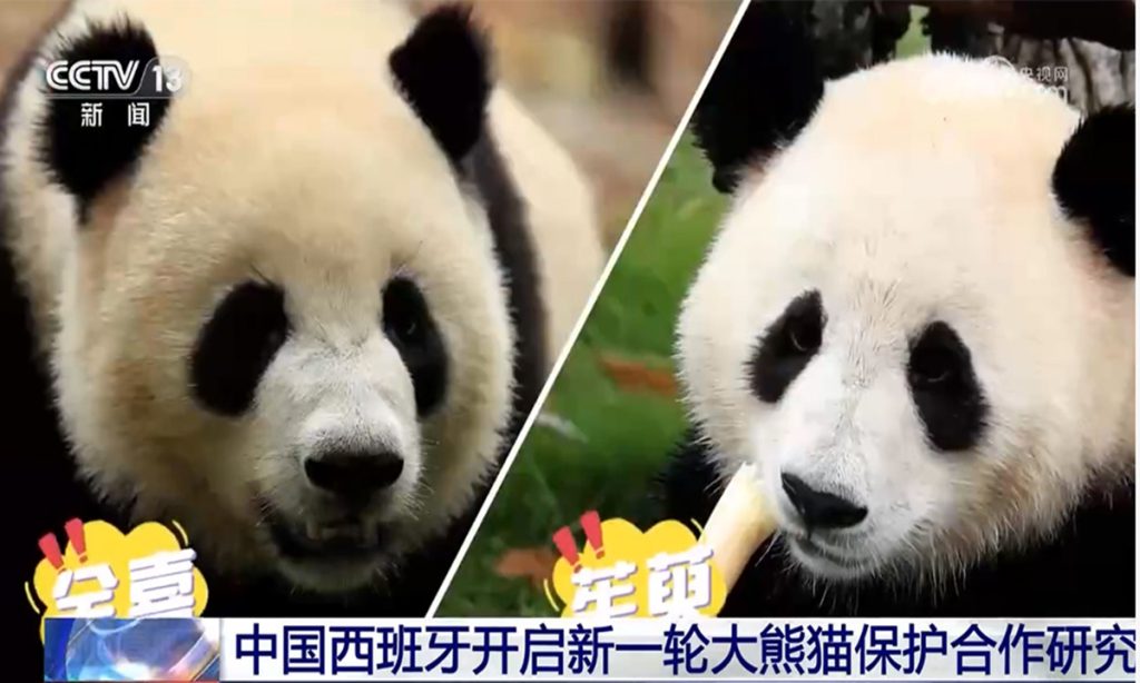
The giant pandas "Jin Xi" and "Zhu Yu" on Monday took off from Chengdu Shuangliu International Airport, bound for Madrid Zoo in Spain to begin a 10-year stay.
Chinese netizens and panda fans have expressed their hope that the two pandas will be well taken care of in Spain. "While abroad, please let the Spanish fans take over the guardianship and take good care of you two babies!" one netizen wrote.
"As the 20th generation of pandas, you two can fly the highest and farthest, opening a new chapter in your panda lives. Stay safe and healthy in a foreign land, and may you spend these long 10 years happily and joyfully!" said another.
"Jin Xi," a male panda, was born on September 1, 2020. "Zhu Yu," a female giant panda, was born on October 25, 2020.
According to Chinese flight tracking platform Feichangzhun, the China Airlines CA3103 cargo plane is responsible for transporting the pandas. The flight took off at 11:24 am on Monday Beijing time, with an estimated flight time of 13 hours and 53 minutes, arriving at Madrid Barajas Airport at 7:20 pm, local time. This is the first pair of pandas to travel to Europe for residency since the second half of 2019.
To ensure the smooth arrival of the two pandas in Spain, both China and Spain have made thorough preparations. The Chinese panda base has isolated and quarantined the two pandas ahead of travel, ensuring dedicated care and regular check-ups.
To ensure comfort and safety during the transportation, the panda base has customized special air transport cages to allow the pandas to move, eat, and rest freely inside the cage. Additionally, the panda base has prepared an ample supply of fresh and high-quality local bamboo and bamboo shoots from Southwest China's Sichuan Province, and arranged for a veterinarian and three specialists to accompany the pandas on the flight to take care of "Jin Xi" and "Zhu Yu's" health and diet.
After arriving at Madrid Zoo, Chinese experts will stay for about three months to accompany the pandas through the quarantine period, complete stress adaptation and behavioral training, and help them quickly adapt to their new living environment.
They will also provide technical guidance and operational training on how to take care of pandas to the zookeepers and veterinarians at Madrid Zoo.
The Madrid Zoo told the Global Times that they will send a press release and videos once the cubs arrive.
Following suggestions of the Chinese expert team, Madrid Zoo has upgraded the panda pavilion in advance, including renovating the cub nursery to meet the needs of panda cub care, and optimized the facility design to cater to the pandas' climbing habits by adding climbing frames, logs, and other play facilities. Those upgraded facilities will be put into use after passing an inspection by the Chinese team.
Spain has achieved the most fruitful results in the cooperative breeding of giant pandas in Europe. Six giant pandas have successfully been given birth in Spain since the cooperation between China and Spain started. The "panda bond" between China and Spain has been maintained for over 40 years.
According to the Madrid Zoo, after arriving in Madrid, the two giant pandas will undergo a one-month quarantine isolation period, and they may meet the public as early as the end of May local time.
Officials, scholars, and business representatives agreed to further promote the development of the brain-machine interface (BMI) technology and fully tap into the broad opportunities of frontier sector at the 2024 Zhongguancun Forum (ZGC Forum) on Friday.
Their statements and comments came at the Brain Computer interface Innovation Development Application Forum, a subforum of the ZGC Forum on Friday, one day after China's first high-performance invasive BMI shine the ZGC Forum, among nine other major technological results.
Xie Yuansheng, an official from the Ministry of Industry and Information Technology (MIIT), said during the subforum that the development of BMI technology will be prioritized as a major future industry, and vowed to enhance policy guidance to the sector.
The MIIT pledged to further promote opening-up and cooperation linked to the BMI technology sector, in order to tap in potential development prospects, said Xie, adding that the innovation capability, industrial integration and regulation system will be further strengthened.
The Brain Computer Interface Industrial Alliance, unveiled 10 cases of industrial innovation in brain-computer interfaces developed by enterprises at the subforum, including cognitive ability and mental health measurement systems, wireless wearable high-speed brain-computer interaction devices, and portable brain-computer interface driving safety intelligent control systems.
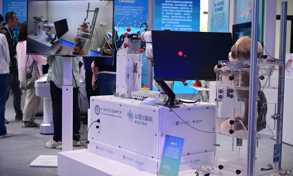
China's self-developed NeuCyber Array BMI System, unveiled on Thursday at the 2024 ZGC Forum, among the 10 cutting-edge technological results, is a high-performance invasive BMI that could be widely applied for medical treatment for patients struggling with neurological and psychiatric brain diseases.
Core high-performance components like high-throughput flexible microelectrodes of the NeuCyber Array BMI System are domestically produced, with performance comparable with the most advanced international levels, Zhao Bing, an engineer with Chinese Institute for Brain Research, said at ZGC Forum.
NeuCyber Array BMI System is China's independently developed and first high-performance invasive BMI, Li Yuan, business development director of NeuCyber NeuroTech (Beijing) Co, the company that developed this BMI brain chip system, told the Global Times on Friday.
The development of BMI technology has broad application prospects, including many with a science fiction flavor, and it is expected that various applications will accelerate landing in near future, contributing to improving the quality of life for the public, Gu Xiaosong, a member of the Chinese Academy of Engineering, said at the sideline of the subforum.
In terms of patent transformation and talent cultivation in the field of BMI technology, China is currently leading the world and outpaced the US. In this regard, China's BMI industry has promising prospects, Gu said.
Research teams from Tsinghua University, Beihang University, and other Chinese universities are among the top in international rankings in BMI sector, Gu noted.
Mohamad Sawan, an academician from the Canadian Academy of Engineering and an Institute of Electrical and Electronics Engineers Fellow, told the Global Times on Friday that China can provide a better environment and cooperation ecosystem for BMI research, as China has "an equation doesn't exist outside."
Sawan noted that the BMI research needs to integrate multiple disciplines together, and universities, academies of science and engineering, institutes for medical engineering in China make collaboration more efficient and faster.
When speaking in which area that BMI technology can benefit more people, Sawan pointed out that some health condition cannot be recovered by drugs or surgery, and it may be applied with BMI equipment for providing economical solution, adding that China has the potential to take the lead in this sector.
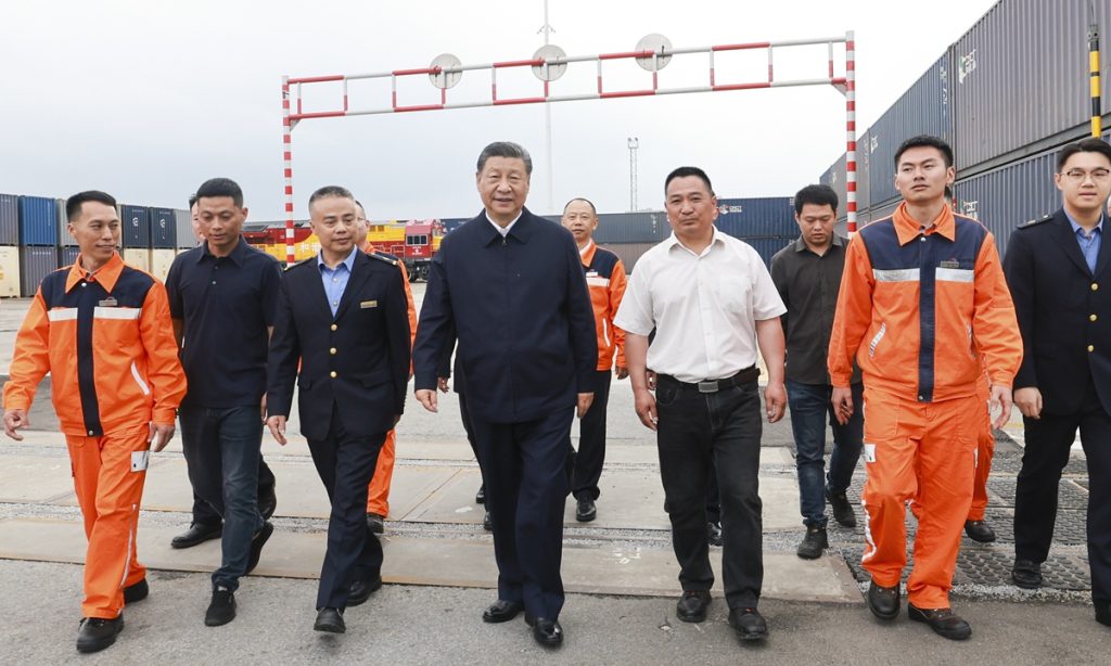
Chinese President Xi Jinping stressed further efforts to usher in a new stage in the development of China's western region featuring well-coordinated environmental conservation, greater openness and high-quality development, when he chaired a symposium on boosting the development of China's western region in the new era in Southwest China's Chongqing Municipality.
Xi, also general secretary of the Communist Party of China Central Committee and chairman of the Central Military Commission, made an inspection tour in Chongqing from Monday to Wednesday. During the visit, he called on Chongqing to further deepen reform and opening up across the board and write its own chapter in advancing Chinese modernization.
The remarks and the inspection tour highlighted the great importance the top leader attaches to the development of the western region, which covers more than 70 percent of China's land area and is home to nearly 30 percent of the country's population. The region plays a critical role in China's pursuit of high-quality development and Chinese modernization, and holds great potential in various areas, economists said on Wednesday.
During the symposium on Tuesday, Xi said China's western region has seen significant achievements on eco-environmental conservation and restoration over the past five years, but the region's development is still facing difficulties and challenges, according to the Xinhua News Agency.
The Chinese President said developing industries that leverage local strengths should be the main focus, adding that efforts are needed to adopt a region-specific approach in developing emerging industries and expedite industrial transformation and upgrade in the western region.
Underscoring the importance attached to greater openness, during the inspection tour of Chongqing, Xi also visited an international logistics hub park to learn about the municipality's efforts in accelerating the development of the New International Land-Sea Trade Corridor in west China, according to Xinhua.
China's western region covers Chongqing, six provinces and five autonomous regions, including Southwest China's Sichuan and Northwest China's Xinjiang Uygur Autonomous Region, accounting for 21.5 percent of China's GDP. Although the region's development has been lagging behind the country's eastern coastal regions, in recent years development has been picking up pace. Between 2019 and 2023, the region's combined GDP expanded form 20.5 trillion yuan ($2.83 trillion) to 26.9 trillion yuan, with an annual average growth rate of 4.9 percent.
"The development in the western region has improved significantly in recent years in terms of infrastructure, industrial upgrade and people's livelihoods," Hu Qimu, a deputy secretary-general of the digital-real economies integration Forum 50, told the Global Times on Wednesday.
In terms of industrial development, the western region has also seen rapid development in recent years. The region is home to nine national level strategic emerging industry clusters in areas such as new materials and biomedicine, and five national level advanced manufacturing clusters such as electronic information and aviation. The region's industrial added value has jumped from 5.8 trillion yuan in 2019 to 8.1 trillion yuan in 2023, according to official data.
Critical role, vast potential
The symposium on Tuesday further highlighted the western region's critical role in China's efforts to bolster both security and development, as well as its enormous potential in areas such as advanced manufacturing and high-level opening-up, economists said.
"The region is tasked not only to boost local industrial development with many industries shift from the east to the west and provide support in resources and other areas, but also to play a bigger role in the country's opening-up efforts," Dong Shaopeng, a senior research fellow at the Chongyang Institute for Financial Studies at Renmin University of China, told the Global Times on Wednesday.
Dong noted that there is still great room for the western region to expand opening-up by bolstering connectivity with countries and regions through land transport corridors.
The western region has developed multiple trade routes with countries and regions in Europe, Central Asia and Southeast Asia in recent years. The New International Land-Sea Trade Corridor, which connects China's western region with Southeast Asian countries, can reach 490 ports in 120 countries and regions as of January 2024, with cargo volume jumping 21 percent year-on-year in 2023, according to official data.
Moreover, the western region also plays a crucial role in the China-Europe Freight Train Express. Over the past five years, 35,000 China-Europe freight trains have been launched in the western region, accounting for 50.5 percent of the national total. Thanks to the greater connectivity, the total import and export volume of the western region reached 3.7 trillion yuan in 2023, an increase of 37 percent from 2019.
Greater connectivity and openness of the western region is of great importance to the country's efforts to expand high-level opening-up and bolster both security and development, economists said.
"Although the capacity of land transport remains relatively low, it has a very high level of security compared to sea transport," Hu said, noting that land transport corridors in the western region can better cope with geopolitical tensions and offer a steady channel for transport of strategic resources under extreme circumstances.
Apart from its great importance in China's long-term security and development, the western region is also crucial in the country's efforts to tackle downward pressure and consolidate the economic recovery in the short term by boosting domestic investment and consumption, economists said.
"Whether it is from the perspective of responding to international situation, or overall coordination of development and security, or the current need to stabilize growth, it is imperative to boost the development of the western region," Hu said, pointing to the huge potential in infrastructure investment and consumption in the region.
As the region's economic development accelerated in recent years, infrastructure construction and consumption has also been expanding steadily. For example, in the first quarter of 2024, Chongqing saw a 4.4-percent growth in fixed-asset investment, with a 16.7-percent growth in industries and 8.5 percent in infrastructure. Retail sales in the municipality also grew by 5.5 percent year-on-year, higher than the national growth rate of 4.7 percent, according to official data.
He Jie, winner of the Beijing Half Marathon, and three African pace setters who appeared to finish as runners-up, have all been stripped of their trophies, medals and prize money, the organizing committee announced on Friday.
The men's champion won the Beijing Half Marathon on April 14 with a time of 1:03:44, just one second ahead of Ethiopian runner Dejene Bikila, and Kenyans Robert Keter and Willy Mnangat, who tied for second place.
The marathon results immediately caused an uproar, as footage showed that He won the race because the three African runners slowed down to let him cross the finish line first.
Amid the ensuing controversy, the organizing committee of the marathon set up an investigation group to find out what happened.
According to the regulations of the event, apart from athletes who apply for the competition by themselves, pace setters can also run in the race but not as competing athletes.
In this event, China Olympic Road Running Company, one of the event organizers, invited a total of 28 Chinese and foreign athletes, including He Jie and four foreign pace setters, at the recommendation of Xtep International Holdings, the sponsor and partner of the sports event.
However, Xtep did not clearly label the pace setters, which led to them being identified as invited athletes who had come to participate in the race.
During the race, the four foreign pace setters wore the same bibs as invited athletes, even though they were supposed to run the marathon as He's pace setters.
One of the four pace setters quit the race soon after it began, but the other three ran all the way ahead of He until the final stretch. They then took the initiative to slow down during the last 2 kilometers. Eventually, He overtook them and won the race.
In accordance with the spirit of fair competition in sports and relevant regulations issued by China's General Administration of Sport, the event organizing committee decided to revoke the competition results for He, Mnangat, Bikila, and Keter, as well as taking back their trophies, medals and prize money. The incident was also reported to the Chinese Athletics Association.
Meanwhile, China Olympic Road Running Company's qualifications for organizing and operating the Beijing Half Marathon have been revoked. It was ordered to issue a public apology and to impose severe punishment on relevant personnel.
In addition, Xtep's partnership with the event has been canceled and it was also ordered to apologize to the public and seriously deal with the personnel held accountable for the incident.
Beijing Sports Competitions Administration and International Exchange Center, the other organizer of the event, was also criticized.
After the punishment decisions were announced, China Olympic Road Running Company apologized in a statement released on Friday, saying that the company expressed deep regret for the negative impact of the incident, and expressed their sincere apology to all the runners and to society.
The punishment decisions were applauded by the public, with many commenting that the individuals and relevant institutions deserve the punishment for profiting through fraudulent means.
Chinese media professional Hu Xijin wrote on Weibo that the relevant individuals and institutions had violated the rules of the competition and deceived the public, going against the spirit of sportsmanship and integrity.
China and the US have recently engaged in intensive dialogues and communications across multiple fields, involving military, financial and economic aspects. Observers believe the current frequent interactions between the world's two largest economies are positively meaningful and conducive to better managing differences between the two sides, and also beneficial for the US to form correct understanding and judgments about China's actions and policies.
However, the most prominent feature of the current China-US relations is that Washington continuously raises demands with China but lacks sincerity in responding to many of China's reasonable requests. Although bilateral relations have relatively stabilized, whether they can progress further remains to be seen, observers said, who also warned to be vigilant against Washington's "tricks," especially in military sector.
Chinese Defense Minister Dong Jun held a video call with U.S. Defense Secretary Lloyd Austin on Tuesday. Dong said the heads of state of China and the United States are committed to stabilizing and improving bilateral relations, stressing that both militaries should serve as a cornerstone for maintaining such stability.
He underscored that the Taiwan question is at the core of China's core interests, which brook no compromise. The Chinese People's Liberation Army stands firm against any activities seeking "Taiwan independence" or external support for such separatist actions, Dong said.
Noting the overall stability in the South China Sea situation, Dong urged the United States to recognize China's firm stance and take practical actions to uphold regional peace, as well as the stability of relations between the two countries and militaries.
Meanwhile, the economic and financial working groups of China and the US held their fourth meeting in Washington DC on Tuesday, with the two sides engaging in "in-depth, pragmatic and constructive" dialogue on how to implement the consensus reached earlier by the leaders of both nations, the macroeconomic situations of both countries and the world, how to achieve balanced growth, and other topics.
The fourth meeting came after the US Treasury Secretary Janet Yellen wrapped up a high-stakes six-day visit to China last week, during which the two countries reached new areas of consensuses in economic and financial fields and also agreed on future meeting arrangements.
From the meeting between the leaders of China and the US in San Francisco last November to their phone call in early April this year, there has been effective communication between the two sides in practical areas. Regardless of whether differences still exist, the communication itself is effective and represents a breakthrough, some Chinese experts said.
Analyzing statements released by the US, Li Haidong, a professor at the China Foreign Affairs University, believes US' sincerity in communication and exchanges with China is insufficient.
"The starting point of their communication is not aimed at moving toward China to better manage differences and resolve conflicts. Instead, the US is motivated by the need to bolster its allies' confidence in how it handles relations with China, demonstrating its capability to keep China-US relations under control. This, in turn, assures its allies to more confidently follow the US in its strategic competition with China," Li told the Global Times on Wednesday.
Vigilance against 'tricks'
The latest talk between the defense chiefs of the two countries, which is a major breakthrough in fully resuming the military-to-military communication, is likely to add possibilities for a future face-to-face meeting at Shangri-La Dialogue in Singapore later once both sides confirm their attendances, some experts said.
But there are still many uncertainties, which are largely linked to the upcoming actions of the US, or whether the US will challenge and break China's bottom line on its core interests.
A US P-8A anti-submarine patrol aircraft flew through the Taiwan Straits and hyped it up publicly on Wednesday. The Chinese People's Liberation Army (PLA) Eastern Theater Command organized warplanes to follow, monitor and deal with the trespassing US aircraft in accordance with the law and regulations, the command's spokesperson said.
The theater command troops are on high alert at all times, resolutely safeguarding national sovereign security and regional peace and stability, the spokesperson added.
In a readout issued by the Pentagon on Tuesday, it reiterated that the US remains committed to "our longstanding one China policy, which is guided by the Taiwan Relations Act, the Three US-China Joint Communiques, and the Six Assurances, and he reaffirmed the importance of peace and stability across the Strait."
Austin also underscored the importance of respect for high seas freedom of navigation guaranteed under international law, especially in the South China Sea, according to the readout.
Philippine and US forces will simulate retaking enemy-occupied islands during joint military drills starting next week in areas facing Taiwan and the South China Sea, Reuters reported on Wednesday.
While military communication between China and the US can be regarded as having fully restored, it's evident from the readouts of both sides that China is well aware of US' ulterior motives and tricks, observers said.
On Lai Ching-te, who has been elected as Taiwan's regional leader in January and will take office in May, I believe that Chinese side is definitely sounding the alarm to the US, which is that the US must fulfill its promise not to support Taiwan independence, Wu Xinbo, director of the Center for American Studies at Fudan University, told the Global Times on Wednesday.
On the South China Sea issue, China's message is also clear that the US should not support the Philippines in its provocations, as such support won't change the overall situation, the expert said. "China and the relevant countries can manage and maintain the overall stability of the South China Sea, and the US' schemes will not succeed," Wu said.
Caution needed
The China-US relationship has not yet fully emerged from its low point, but it is relatively stable, or rather, "there are still pitfalls ahead, but both sides have lights to guide them," Lü Xiang, a research fellow at the Chinese Academy of Social Sciences, told the Global Times on Wednesday.
"Both parties have flashlights in hand, so they are unlikely to fall into a pit. This is the current situation, but there is still the possibility of accidentally stepping into a pit, so both sides should remain cautious," Lü said.
Despite that the economic and financial working groups of China and the US had substantial dialogue in Washington DC, the US Trade Representative Katherine Tai is reportedly going to tell lawmakers that the Biden administration is "taking a serious look" at US trade defense tools to deal with threats posed by China's trade and economic policies, including a review of Trump-era tariffs on Chinese imports.
"The core issue is that the US no longer adheres to or follows its own established rule, arbitrarily causing the current chaos in the global economic and international trade sectors," Gao Lingyun, an expert at the Chinese Academy of Social Sciences, told the Global Times on Wednesday.
It's also difficult to predict whether tensions in the economic and trade sectors between the two countries will ease following the latest interactions, but the two sides could focus on areas of mutual benefits before moving on to topics where there are greater disagreements.
While interactions between China and the US have increased, whether the relationship between the two countries can progress beyond stability is yet to be seen, observers said.
"It seems the US is more concerned with maintaining stability, ensuring that no major issues arise. In contrast, China hopes to address some mutual concerns while maintaining stability, to promote an improvement in bilateral relations," Wu said.
To improve, issues need to be resolved and progress made. However, the US currently shows little interest in moving forward, including in military relations and it is more focused on preventing any accidents or crises than on engaging in extensive military exchanges, the expert said.
The current dynamic is characterized by Washington continuously pressuring China and raising demands, with a lack of a positive and sincere response to China's legitimate concerns. "In this context, Blinken's upcoming visit to China won't be an easy task, and we should not expect significant outcomes," Wu added.
Responding to "concerns" from a pro-US political figure in the Solomon Islands over China's growing influence ahead of the nation's elections, a Chinese Foreign Ministry spokesperson said on Monday that China adheres to non-interference principle in other countries' internal affairs, and advocates prioritizing development while respecting the Pacific nation's autonomy.
Experts noted that the "concerns" appear more like a slogan for the US with the aim of suppressing China's interests in the South Pacific region. Since establishing diplomatic relations, China's cooperation with and assistance to the Solomon Islands have been conducted under the principle of mutual respect without any political conditions, they stressed.
Daniel Suidani, a major rival of the incumbent Prime Minister Manasseh Sogavare and the former premier of the most populous island Malaita, said in a recent interview that China's presence in his nation is "alarming," noting that the upcoming elections could further "entrench Beijing's foothold."
One of the few provincial leaders in the country to refuse cooperation with China, Suidani claimed that he fears the assistance could one day "come with strings attached," while accusing China of meddling in the Solomon Islands' elections without providing evidence.
In response to the allegations, Chinese Foreign Ministry spokesperson Lin Jian said at Monday's press briefing that China has always adhered to the principle of non-interference in other countries' internal affairs and supports the people of the Solomon Islands in choosing a development path that suits their national conditions.
China advocates that all countries, in developing relations with Pacific island countries, should respect their autonomy, prioritize development, and promote inclusiveness, Lin noted.
"In fact, Suidani's province of Malaita and he himself has long had a close bond with the separatist authorities of Taiwan island. In addition, he is heavily influenced by the US, which has led to a bias against China," Chen Hong, executive director at the Asia Pacific Studies Centre of East China Normal University, told the Global Times on Monday.
According to media reports, Suidani has previously signaled willingness to re-establish ties with traditional security partners including Australia and the US.
Washington has been trying to manipulate the politics of the Solomon Islands since Sogavare took office, Chen noted, by pressuring the current government through various means such as media and public opinion, while at the same time cultivating individuals within opposition parties to influence the country's domestic and foreign affairs.
Another of Suidani's aims is to differentiate himself from the current government by emphasizing his stance on China, so as to enhance his political profile and gain support ahead of the election, observers said.
Voters from among 700,000 people spread over the more than 900 islands in the country will go to the polls on Wednesday to elect 50 lawmakers from 334 candidates. The 50 newly elected lawmakers then decide which of them will become prime minister.
Sogavare is seeking a second consecutive term, AP reported on Monday.
Western media, in line with US strategy, has spared no efforts to exploit this election as a chance to disrupt the relations between China and the Solomon Islands, claiming that the election could impact China's cooperation with the Pacific nation.
Shrugging off this hype, Chinese experts said that the tangible benefits China has brought to local people are clear, whether it's in infrastructure development or in comprehensive economic cooperation.
"Attempts by certain politicians to reject China's partnership ultimately goes against the interests and needs of its people, and are doomed to fail," Chen said.
Several previous cases have also proven that while some politicians may exploit anti-China rhetoric during their countries' elections, once in power, they quickly came to realize the benefits of cooperating with China and soon switch to a prudent and pragmatic approach in dealing with China, Chen noted.
Since severing ties with the island of Taiwan and establishing diplomatic relations with Beijing in 2019, the Solomon Islands has signed a security pact with China in 2022, and inked a comprehensive strategic partnership featuring mutual respect and common development for a new era in 2023 during Sogavare's visit to Beijing.
Research unveiled during the inaugural China Humanoid Robot Industry Conference in Beijing from April 9 to 10 indicates that the global humanoid robot industry is entering a golden era, poised for sustained growth. The report predicted that the Chinese humanoid robot market will surpass 10 billion yuan, reaching 10.47 billion yuan ($1.45 billion) by 2026, and is anticipated to soar to 119 billion yuan by 2030.
The booming market is seemingly telling people that intelligent humanoid robots that can simulate human thinking and consciousness, as depicted in films like Ex Machina and A.I. Artificial Intelligence, are really getting closer to reality.
The rapid development of AI technology has played a crucial role in the advancement of humanoid robots. As NVIDIA CEO Jensen Huang said at the 2024 GPU Technology Conference (GTC) in March, "Building foundation models for general humanoid robots is one of the most exciting problems to solve in AI today."
In return, the advancement of humanoid robots is viewed as a major milestone in the AI era, pushing the boundaries of AI research. Numerous Chinese experts and industry observers consider humanoid robots a breakthrough for the "AI Plus" initiative aimed at fostering innovative development in the digital economy, as promoted during the two sessions in March.
Golden age along with AI
Humanoid robots are dubbed "humanoid" because they are designed to emulate and potentially surpass human capabilities in form, function, behavior, and even cognitive processes, Zhang Rui, founder and executive director of the Beijing Ironman Technology in Beijing, told the Global Times.
"Without the need for massive changes to the existing environment, humanoid robots can seamlessly integrate into various scenarios, using their flexible and dynamic execution capabilities to meet complex and changing task requirements. Furthermore, their human-like characteristics enable them to easily manipulate human tools, further expanding their application areas," Zhang said.
Therefore, humanoid robots are not only a symbol of technological progress but also a significant force driving future social development, he noted.
Humanoid robots have been widely applied in various industries, with the aerospace sector being one of the most prominent, according to Zhang. Several countries including the US, Russia and China, have been deeply researching the application of humanoid robots in the aerospace field. These robots are mainly used to replace humans in performing dangerous and complex operations, ensuring the safety of astronauts and improving the efficiency and success rate of space missions.
Other major application areas of humanoid robots is in border defense and lights-out factories, or smart factories.
"The continuous innovation and breakthroughs in AI technology in recent years have indeed provided humanoid robots with more powerful perception, decision-making, and execution capabilities. This allows humanoid robots to more accurately understand human language, recognize environmental information, and make more reasonable decisions and actions," Zhang said.
In the future, Zhang expects humanoid robots to have enormous potential in areas such as general hardware execution, dynamic adaptation and environmental integration.
How far is AGI?
The thriving progress of humanoid robots is drawing increasing attention from international tech giants. On March 18, nine humanoid robots were unveiled at NVIDIA's 2024 GTC. Tesla is also actively working on a humanoid robot named Optimus, and OpenAI, Microsoft, and Amazon founder Jeff Bezos have made substantial investments in humanoid robot startup Figure AI. Additionally, Agility Robotics, backed by Amazon, has established the world's first large-scale humanoid robot production factory in Oregon, the US, capable of producing 10,000 two-legged robots annually.
A group of innovative and competitive Chinese companies have also emerged in the field of humanoid robots with the increasing emphasis and investment in robot technology in China, leading to significant progress in the Chinese humanoid robot industry.
In March, the Beijing humanoid robot innovation center announced that it would soon release the first generation of a universal open humanoid robot body.
Among the nine robots showcased at the NVIDIA 2024 GTC, two were developed by Chinese companies, namely H1 from Hangzhou Unitree Robotics and PX5 from Xiaopeng Pengxing.
The Global Times learned from Unitree Robotics that H1 is a full-size humanoid robot capable of running, equipped with 360 panoramic depth perception. Currently, it can reach a speed of 3.3 meters per second, setting a world record for full-size electric humanoid robots, with a potential speed of up to 5 meters per second.
This robot boasts highly advanced full-body dynamic coordination capabilities, enabling it to dance in groups and execute backflips. As a result, NVIDIA opted to partner with Yushu to collectively propel the global advancement of AI robots. According to the company's response to the Global Times, NVIDIA, a frontrunner in GPU and AI chip technology, furnishes Yushu's robots with robust computing capabilities and comprehensive support in deep learning technology.
However, overall, the deep integration of AI and humanoid robots still faces significant challenges.
Zhang believes hardware challenges are a crucial obstacle. While we can achieve various complex functions and performance at the algorithm level, it is often difficult to achieve the desired output power and efficiency in actual robot hardware, he said.
This is mainly due to the numerous technical details and engineering challenges involved in hardware design and manufacturing, requiring continuous optimization and improvement, Zhang explained.
On the other hand, the current progress of AI technology is mainly limited to deepening and innovating at the logical level, with insufficient breakthroughs in thinking and emotional aspects. While the form of robots is malleable, the "spirit" of their internal thinking and emotions is still an unexplored frontier. Zhang believes it will take another 5-10 years to achieve a 70 percent similarity with human emotions.
At an economic forum held at Stanford University in March, Jensen Huang predicted that a general artificial intelligence that can pass human tests, or "human-like" artificial general intelligence (AGI, capable of performing all human intelligent behaviors), is likely to appear within five years. However, Huang also pointed out that achieving this goal is not without difficulties, as scientists still lack a unified definition of how the human mind operates, making it challenging for engineers to achieve the goal.
On April 8, Elon Musk said during a livestreamed interview on X that AI that is smarter than any one human will probably come around by the end of next year. Last year, he predicted that humans would "fully" achieve general artificial intelligence by 2029.
Some experts believe that with the continuous advancement of chips and algorithms, AI may eventually surpass human intelligence. However, Liu Wei, director of the human-machine interaction and cognitive engineering laboratory of the Beijing University of Posts and Telecommunications, pointed out that AGI may be a false proposition. This is not because current AI systems have not reached the level of general intelligence, but because AI fundamentally perform and learn like humans.
The development of AGI faces three major bottlenecks: technical, biological, and social. The technical bottleneck lies in the need for AI systems to have higher computing power, more advanced algorithms, and more efficient data processing methods to achieve more complex and intelligent functions. The biological bottleneck mainly stems from our limited understanding of the cognitive capabilities and operation mechanisms of the human brain, requiring deeper research in neuroscience and cognition to achieve similar levels of intelligence. The social bottleneck includes the integration of AI systems with human society, such as cultural differences, ethical issues, privacy protection, etc., all of which are crucial factors affecting the development of AI, according to Liu.
"To overcome these bottlenecks, interdisciplinary cooperation and continuous innovation efforts are needed. Only by making breakthroughs in technology, biology and society can AGI move towards more mature and comprehensive development, but it remains extremely difficult, perhaps impossible after all," Liu said.
Safety and ethics concerns
In November 2023, the Ministry of Industry and Information Technology of China issued the guiding opinions on the innovation and development of humanoid robots, proposing to establish a preliminary innovation system for humanoid robots by 2025.
The document predicted that, by 2025, key technologies such as those related to robots' "brain, cerebellum, limbs" will achieve breakthroughs, ensuring safe and effective supply of core components. The whole machine products will reach international advanced levels, achieve mass production, and be demonstrated in special, manufacturing, and civil service scenarios, exploring effective governance mechanisms and means. By 2027, China's comprehensive strength in humanoid robots will reach world-class levels, becoming an important new engine for economic growth.
The development of the humanoid robot industry, driven by both technology and policy, has entered the fast lane of development, but it also faces challenges in safety, social ethics, and legal norms. Some experts point out that in scenarios requiring close contact, such as in elderly care or assistance, where physical contact is needed, the safety and risk issues of humanoid robots cannot be underestimated. More research is needed by the industry and relevant regulatory authorities on the technical and application scenarios.
Zhang believes that when unexpected situations occur with humanoid robots in a home environment, the lack of clear legal definitions makes it difficult to determine responsibility and protect rights. Additionally, there is currently no mandatory system for certifying the eligibility of humanoid robot products, making it challenging to distribute products in large quantities and limiting the widespread application of humanoid robots. Therefore, it is essential to address and resolve these legal gaps as soon as possible to ensure the healthy and orderly advancement of humanoid robots in the future.
However, he firmly believes that with the continuous progress of technology and the gradual improvement of regulations, humanoid robots will demonstrate their unique charm in more fields, contributing more to the development of human society.
In 2023, the Chinese Ministry of Science and Technology, together with the Ministry of Education, the Ministry of Industry and Information Technology, and 10 other departments, issued the trial ethical review measures for science and technology.
According to the measures, carrying out scientific and technological activities should adhere to the unity of promoting innovation and preventing risks. It involves objectively evaluating and prudently handling uncertainties and risks of technological applications, following the principles of enhancing human well-being, respecting the rights of life, upholding fairness and justice, reasonably controlling risks, and maintaining openness and transparency in science and technology ethics. It is essential to comply with the Constitution, laws, and regulations of China, relevant provisions, and ethical norms of science and technology.
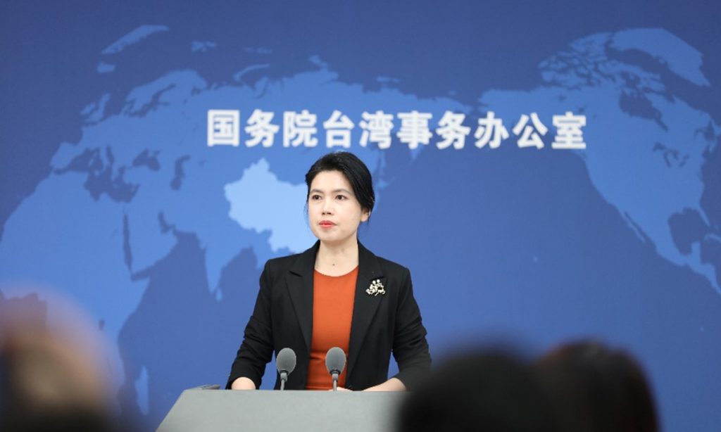
The Chinese mainland continues to support cross-Straits exchanges and cooperation in various fields despite the Democratic Progressive Party (DPP) authorities consistently obstructing exchanges. Their so-called “goodwill” to enhance exchanges has not been seen, Zhu Fenglian, spokesperson for the Taiwan Affairs Office of the State Council told a press conference on Wednesday.
Zhu’s remarks came after reports that Taiwan's transportation officials recently claimed that due to the decrease in mainland tourists, the target number of tourist trips to Taiwan this year would decrease from 12 million to 10 million. And there is public outcry within the Taiwan island urging the DPP authorities to seek solutions and not allow Taiwan's tourism industry to become a political sacrifice.
“The DPP claim that they have been showing goodwill since last year, but what we see is that there has still been no formal opening for mainland residents to visit the Taiwan island, nor has the ban on Taiwan residents participating in group tours to the mainland been lifted. Where is the so-called goodwill? We haven't seen it,” said Zhu.
During the Wednesday press conference, Zhu also criticized the DPP authorities for obstructing exchanges between young people.
Former chairman of the Chinese Kuomintang party (KMT) Ma Ying-jeou is leading a Taiwan youth delegation to visit the mainland this week, including Guangdong, Shaanxi, Beijing and other places to trace their roots and exchange experiences.
Zhu said that the foundation of cross-Strait relations lies in the people, and the driving force comes from the people. She hopes that young people on both sides of the Taiwan Straits can bravely shoulder responsibilities, unite in friendship, and work together.
The mainland has consistently advocated and actively promoted cross-Straits youth exchanges, and will continue to create favorable conditions for mutual learning and exchange between young people, and provide more conveniences for Taiwan youth in studying, working, starting businesses, and living on the mainland, Zhu said.
Mr Ma has led young people from the Taiwan island to visit and conduct exchanges on the mainland twice, and invited faculty and students from five mainland universities to visit the island, making significant contributions to promoting cross-Straits youth exchanges. “We fully acknowledge and appreciate this,” said Zhu.
The main obstacle to cross-Straits youth exchanges at present lies in the obstruction and restrictions imposed by the DPP authorities, Zhu said, noting that they will work with people of both sides of the Taiwan Strait to actively carry out various activities to promote youth exchanges, allowing more young people from both sides to get to know each other, work together, and inject youthful vitality into the continuous development of cross-Straits relations.
While visiting the former residence of Sun Yat-sen, a great national hero, great patriot and great forerunner of China's democratic revolution, in Guangdong, Ma called on the two sides of the Taiwan Straits to work together and “avoid war.”
Zhu said that the peaceful development of cross-Straits relations is the correct path for safeguarding peace, promoting common development, and benefiting compatriots on both sides. People on both sides of the Straits should work together to fulfill the Chinese dream together, and shoulder the responsibility of national rejuvenation.
The DPP authorities persist stubbornly in their separatist "Taiwan independence" stance and collude with external forces to continuously provoke separatism, which is the root cause of tension and instability in the Taiwan Straits.
We firmly oppose "Taiwan independence" and will not leave any room for separatist activities. The DPP authorities collude with external forces to provoke "independence" and undermine peace in the Taiwan Straits, damaging the interests and well-being of Taiwan compatriots, which is truly detested by the people of Taiwan, Zhu said.