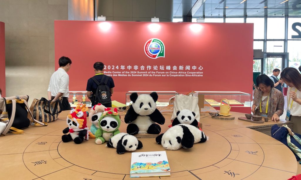
In one exhibition corner, several guests from Chad, Nigeria and Egypt carefully explore traditional cloisonne techniques by painting some cultural products. Nearby, some guests are fully immersed in an 8k ultra HD video experience, which is taking them on a virtual journey from the Central Axis of Beijing to the majestic wildlife migrations of Africa.
This scene took place at the China National Convention Center, the venue for the 2024 Summit of the Forum on China-Africa Cooperation (FOCAC), on Thursday.
During the summit, the newly released China-Africa Cooperation Beijing Action Plan (2025-27) highlighted people-to-people exchanges in terms of culture, tourism and sports.
The plan noted that the two sides will jointly implement the Global Civilization Initiative (GCI), increase engagement and cooperation in areas such as education, science and technology, health, tourism and media, strengthen cultural exchanges, and enhance people-to-people ties, with the aim of cementing the social foundation for friendship between China and Africa.
Many guests from countries including Zimbabwe, Mali and Uganda told the Global Times that FOCAC has consistently been a platform that fosters deeper cooperation and cultural exchanges between China and Africa, particularly in areas critical to sustainable development.
On Tuesday, China signed a number of joint statements with many African countries such as Zimbabwe, Nigeria and Uganda.
Tungamirai Eric Mupona, vice chairman of the China Zimbabwe Exchange Center, told the Global Times that the summit can be seen as a concrete manifestation of China's active implementation of the GCI. He highlighted China's support for Zimbabwe's educational infrastructure and the preservation of its traditional culture.
Mupona also noted the significance of a memorandum signed between the two countries' national television networks and media, which is expected to further promote people-to-people exchanges.
Melha Rout Biel, executive director of the Institute for Strategic and Policy Studies from South Sudan, told the Global Times that South Sudan is home to over 60 distinct ethnic groups, each with their own unique culture and traditions, much like China's 56 ethnic groups. This diversity forms an essential foundation for cultural exchanges between the two countries.
"We are all human beings, so you can learn from others. Cultural interaction brings people closer together," Biel said, adding that South Sudan's rich tradition of arts and crafts offers China a window into African culture and highlighting the potential for mutual enrichment through such exchanges.
Dahlia A. Ducreay, director of the International Department from the Silk Road Youth Forum, told the Global Times that China and Africa can build a partnership that is not only strong but also "just and sustainable." "I would like to emphasize the importance of ensuring that development initiatives under FOCAC and the Belt and Road Initiative are not only economically beneficial but also socially empowering."
The consulates general of Spain, Germany, the Netherlands, Luxembourg, and Poland in Shanghai and the European Union jointly launched the EU Jazz Month from September 6 to 29, creating good, healthy and happy vibes for music lovers.
Music bands, including Marco Mezquida - Tornado from Spain, Malstrom Live Concert from Germany, Fingerprint featuring Shirma Rouse from the Netherlands, Greg Lamy Trio from Luxembourg and Zk Collaboration from Poland, are in the spotlight over the month of September.
From September 6 to 7, Spanish jazz pianist Marco Mezquida, along with his musical partners, bassist Masa Kamaguchi and drummer Ramon Prats, offered a wonderful rendition of the album "Tornado."
On September 8, German Malstrom Live Concert was staged as part of the EU Jazz Month.
Based on jazz, their music blended contemporary influences such as free improvisation, metal, rock, and electronica, with a strong sensory impact and ferocious performance.
Rahmat Allah Mohamed Osman, the permanent representative of the African Union (AU) to China, highlighted in a recent interview with Global Times that the 2024 Summit of the Forum on China-Africa Cooperation (FOCAC) that just concluded in Beijing is significant for China-Africa relations. He believes that China and Africa have mutually beneficial needs, and expresses hopes for the implementation of more policies in China's support in agriculture, industry, and talent development to Africa.
The AU is the first regional international organization to sign a cooperation plan with China-proposed Belt and Road Initiative (BRI) and establish a working coordination mechanism. In Osman's view, China-Africa cooperation can effectively promote the realization of Africa's development blueprint "Agenda 2063" and accelerate industrialization in Africa.
In 2018, the AU established a representative office in China, with Osman, as the first AU permanent representative to China, having witnessed the upgrading of China-Africa relations. In 2023, China launched the Initiative on Supporting Africa's Industrialization, and implemented the Plan for China Supporting Africa's Agricultural Modernization and the Plan for China-Africa Cooperation on Talent Development under the framework of the FOCAC to support Africa's development and prosperity with concrete steps.
According to Osman, these initiatives cover areas that are urgently needed for Africa's modernization efforts and will help African countries enhance their development capabilities.
Industrialization is the necessary path for the African continent to achieve development and is an important cornerstone for Africa's prosperity and strength. Osman believes that deepening cooperation between Africa and China will strongly promote the realization of the AU's Agenda 2063 and accelerate the industrialization process in Africa.
African countries have a significant demographic advantage, with a high proportion of young people, and cooperation between Africa and China in talent development will help to unleash the demographic dividend of African nations, he said.
Osman noted that China and Africa are making progress to heighten trade, with Kenyan flowers and avocados already making their way into the Chinese market, and Ethiopian and Rwandan coffee being enjoyed by Chinese consumers. He believes that trade between China and African countries still has great potential for growth.
The envoy criticized the Western narrative of China engaging in neo-colonialism in Africa. He stated that African countries share many common concerns with China, which has always listened to Africa's voice and provided steadfast support. Historically, Western colonizers plundered Africa's resources and showed indifference to local construction and development in Africa.
"This [narrative of China engaging in neo-colonialism in Africa] is ridiculous and it's not acceptable for us at all," the envoy told the Global Times.
In the harsh, oxygen-thin environment of the Xizang Autonomous Region in Southwest China, where life on the "roof of the world" meets daily challenges, the resilience and unity of the Chinese people have been tested and proven over the last three decades.
Since 1994, approximately 12,000 officials have been dispatched to Xizang through the paired-up assistance mechanism. Among them, Party members have formed the backbone of this mission, which has been sustained across generations.
They are comprised of doctors who brave the cold and lack of oxygen to bring health and hope to remote villages; teachers who cross mountains and rivers to impart knowledge and ignite the dreams of children; engineers who carve roads through treacherous terrain, connecting isolated communities to the broader world; and officials who leave their homes and families behind to dedicate themselves to the development of Xizang, embodying the spirit of perseverance and dedication.
As 2024 marks the 30th anniversary of these dedicated efforts in aiding Xizang, the Global Times has interviewed aid teams from across the region to delve into their inspiring stories.
Long-lasting spirit
Li Manwan has never regretted her decision.
A doctor from Changde, Central China's Hunan Province, Li first arrived at the Lhunze County People's Hospital in Shannan Prefecture, southeastern Xizang in August 2021. After completing her initial year-and-a-half aid mission in Xizang, she applied to join the second batch of medical workers in March 2023, continuing her service in Lhunze to fulfill her medical mission.
Lhunze sits at an average altitude of 3,800 meters and is known for its harsh, windy conditions. Initially, Li overcame the discomfort caused by altitude sickness and took full responsibility for clinical teaching, outpatient services, and surgeries in the hospital's obstetrics and gynecology department.
Within just two years, the obstetrics and gynecology department developed into a comprehensive clinical unit, integrating gynecology, obstetrics, family planning, and health care, with the capability of handling emergency care for pregnant women and newborns.
Li has excelled not only in improving medical techniques, but also in team building.
Over the last three years, Li has also led her team to complete free screenings for the "two cancers" across all 11 townships in the county, serving a total of 1,066 people.
"Lhunze is my second home, and the obstetrics and gynecology department is like a child I have nurtured. I want to continue guiding it forward," Li told the Global Times.
In northern Xizang, a hospital located at an altitude of 4,500 meters has become a shelter for the local people, granting them access to guaranteed medical services.
The Nagqu People's Hospital faced challenges such as its remote location and harsh climate, as well as trouble attracting and retaining highly skilled medical professionals. Nagqu is also the highest-altitude prefecture-level city in the country, with the harshest environmental conditions for local residents. The average elevation in the city is 4,500 meters, and the oxygen content in the air during the summer is only 58 percent of that at sea level. The annual average temperature ranges from -0.9 C to -3.3 C.
Since 2015, Liaoning Province has dispatched a total of 116 experts to assist Xizang, providing strong support for the high-quality development of medical services in Nagqu. Additionally, Liaoning Province has invested over 20 million yuan ($2.7 million) in the purchase of equipment such as magnetic resonance imaging and telemedicine platforms to aid in the hospital's development, the Global Times learned from local government.
Currently, critical care units for maternal and child health, pediatric critical care, a high-altitude medical research center, and emergency rescue ave all been established.
"In the past, women from local herding communities did not have the habit of giving birth in hospitals, but now people are more willing to come to the hospital because it is safer, more reliable, and more hygienic. People also have more faith in the hospital," Zhao Yi, director of the obstetrics and gynecology department at the Nagqu People's Hospital, who comes from Liaoning, told the Global Times. Better life, better future
Some of China's most economically developed regions are also bringing their advanced experiences, skills, and concepts in development to the most remote areas of Xizang.
Medog county, the last county in China to be connected to the national road system, is nestled deep in the southeastern Himalayas. The roaring Yarlung Zangbo River makes a dramatic turn at the Guoguotang Bend, creating abundant hydropower resources in the area.
Standing at the iconic Guoguotang viewing platform, one can look down at a beautiful tea plantation, shaped like a horseshoe, imprinted on the nearby hillside, blending in with the majestic Yarlung Zangbo River Valley.
Today, tea has become the "golden leaf" that enriches the lives of the people of Medog.
Since the first tea bush was planted in a trial in 2015, Medog's tea gardens have spread across the high mountains and deep valleys. During the tea-picking season, farmers are seen constantly moving about, and the fragrance of tea fills the plateau air.
The county has now established 103 high-altitude organic tea gardens, with a total tea plantation area of 19,000 mu (approximately 1,267 hectares). Currently, there are six tea processing enterprises, and in 2023, 5.25 million kilograms of fresh tea leaves were harvested, increasing the income of the local people by 5.13 million yuan ($700,000), according to the Medog government.
This success is a result of aid efforts from Foshan, South China's Guangdong Province. Since 2013, Foshan has sent four groups of 189 officials and professionals to aid Xizang, investing 570 million yuan ($80 million) in aid funds, and completing 89 construction projects. Additionally, they provided 77 million yuan in extra-budgetary funds for five more projects, contributing significantly to the tremendous changes in Medog since the opening of the Medog Highway 10 years ago.
With a decade of infrastructure development and tourism promotion, Medog has now become one of the most popular tourist destinations in Xizang. In 2023, the county welcomed 42,000 tourists, generating over 200 million yuan in revenue, the Global Times learned from Medog publicity department.
Similarly, Yadong county, also located on the border, has benefited from the development of tourism brought about by aid teams.
Pangda village is a model well-off border village supported by Shanghai's aid. Thanks to this support, 99 households with nearly 500 residents relocated from a village at an altitude of 4,630 meters to Pangda at around 2,000 meters, where they have started a new life.
In addition to economic development, Shanghai has also brought advanced educational resources to Yadong.
In recent years, Putuo district's education system has dispatched numerous outstanding officials and teachers to Xizang.
These educators have embedded Shanghai's advanced teaching concepts and methods into the schools they assist, engaging in deep exchanges with local teachers, growing together, and becoming "golden seeds" that foster the reform and development of local education. Lasting legacy
Among the many pairing-up assistance programs for Xizang, the artistic support efforts of a talent team from East China's Fujian Province are also impressive.
Thangka, a scroll painting framed with colored satin, is the most representative folk religious art form of the Tibetan ethnic group. Lacquer painting is a traditional painting form that uses natural lacquer as the main material. Fujian lacquer painting is one of the important lacquer painting schools in China. Under the efforts of the Fujian Aid-Xizang Work Team, they merged to produce a new art form - Thangka lacquer painting.
Since 2018, the Fujian Provincial Museum of Art has explored innovative art poverty alleviation models and has successively sent professional personnel and local lacquer artists to hold nine Thangka painting training sessions in Qamdo, eastern Xizang, and Fujian, teaching over 160 Thangka painters the art of lacquer painting and creating more than 300 Thangka lacquer painting works.
"The technique of Fuzhou bodiless lacquerware and Thangka art are both listed as the first batch of national intangible heritage. Through paired support, these two ancient and locally distinctive art forms have achieved organic integration," Yu Zheng, a representative inheritor of Fuzhou bodiless lacquerware technique, told the Global Times.
Yu went to Qamdo in August 2023 for a month-long course on Thangka painting techniques.
"When we came to Qamdo, the collision of these two art forms brought about an expansion of the local Thangka painters' creativity, ushering these Tibetan artists to a broader stage," said Yu.
In 2019, Thangka lacquer painting works made their international debut at the second China International Import Expo. Since then, these art pieces have appeared at important international cultural exchange platforms such as the Cross-Strait Cultural Industry Expo and the China International Copyright Expo.
In June 2020, nearly 400,000 netizens watched the process of creating Thangka lacquer paintings streamed online.
Fujian has several active lacquer art creation and operation groups in China. These professional technicians and lacquer artists have conducted Thangka lacquer painting training in Xizang, helping local Tibetan artists boost the visibility and sales of their artworks through the fusion of painting techniques, bringing them more income, Yu said.
In 2022, under the guidance of the Fujian talent team, the Qamdo Vocational and Technical College established a major in lacquer painting to cultivate composite talents in this kind of painting.
Yu emphasized that the training of Thangka lacquer painting techniques offers not only technical skills and cultural preservation, but also a mutually beneficial cultural exchange and a deep emotional connection between people from different places. This vibrant artistic innovation continues to attract more and more people.
Located in South Asia, India is China’s close neighbor, yet for many Chinese people, the country remains both intriguing and unfamiliar. Many social media bloggers often share various “incredible” experiences they encounter in India: Asia’s largest slum, the daring and reckless act of “train surfing,” and the “clean and hygienic” street food specialties, among others. While the lively streets and aroma of curry represent a unique side of Indian culture, Mi (pseudonym), a Chinese woman who has lived in India for nearly 20 years, has witnessed more of the country’s changes and its complex “duality.” The Global Times invites Mi to share her perspective on India with our readers. Bumpy roads
Our impression of a strange city often begins at the airport or train station. In 2006, I flew directly from Beijing to New Delhi, the capital of India. As soon as I got off the plane, I was hit by a strong sensory overload: the complex aroma of spices filled the air; the customs officer’s accent made it hard for me to tell whether he was speaking Hindi or Indian English, no matter how carefully I listened; a little sunlight filtered through the thick grease on the glass windows, making it difficult to discern whether it was sunny or cloudy outside.
However, the situation quickly improved. In 2010, the opening of the new Terminal 3 at Indira Gandhi International Airport made it one of the largest and most important airports in South Asia, and it ranked among the world's largest modern airports. The airport's design is rich in elements of India's diverse culture, leaving a deep impression on travelers from all over the world.
When it comes to transportation in India, most people might think of trains packed with passengers. However, the subways and light rail systems in Indian cities are also clean, quiet, and well-equipped, with dedicated compartments for women. Riding the Indian subway in the summer is indeed a pleasant experience: the air conditioning is sufficient, the cars are bright and clean, and some stations even have cafes and convenience stores.
According to a report by Indian media outlet Mint, India’s Union Minister Hardeep Singh Puri said, “Today we have 945 kilometers of metro system functioning in the country, and we have another 1,000 under construction. This will be done in the next two and a half years, and we will have the world's second-largest urban transport metro.”
However, above this rapidly expanding subway network, the streets are a chaotic mix of cars, motorcycles, auto-rickshaws, and pedestrians, often encountering cows, dogs, camels, horse-drawn carts, and elephants. The sound of honking horns and bustling crowds is ever-present. As for highways, India has had them for 20 years, but this "high-speed" is not the same as what one might expect.
In 2006, when a group of us traveled to the Taj Mahal in Uttar Pradesh, I fell asleep as soon as I got in the car. After about an hour, I woke up to see cows, dogs, camels, tractors, and pedestrians still crossing the road, and I asked, “Why haven't we gotten on the highway yet?” Other passengers burst into laughter, saying, "This is the highway!"
Now, India finally has a real expressway. Known as the "Golden Quadrilateral," a network of four national highways that connects New Delhi, Mumbai, Kolkata, and Chennai, spanning approximately 5,800 kilometers. The travel time for the route from New Delhi to the Taj Mahal, which is about 200 kilometers, has been reduced from 5 hours to just 3 hours, with a toll fee of 470 rupees ($5.6).
Looking back 20 years ago, New Delhi had only one relatively decent shopping mall called Ansal Plaza, which was a two-story building. The more upscale shopping street was Connaught Place in the city center, built by former British colonizers. At its center is a circular large park surrounded by white buildings, all two stories high, housing shops, restaurants, cafes, and more. For most people, however, community markets were the preferred shopping destinations. Along a street, there were single-story shops on both sides, which looked like abandoned warehouses from the outside, and the interior arrangements were chaotic and cramped.
Things have improved significantly now. Large shopping malls, supermarkets, and convenience stores are everywhere in New Delhi, and people’s lives have benefited from better infrastructure. However, the situation of pothole-ridden streets and litter-strewn areas in Indian cities have not changed. An Indian friend joked that in India, even if you drive a million-dollar luxury car, you still are forced to experience the bumps. I recall that just before the local elections, the streets in my community were hastily repaired. Although they looked new, the quality of the construction was concerning, and not long after the elections, they returned to their original state. The locals lamented to me, "the government won't make investments without returns."
It wasn't until the 2010 Commonwealth Games and last year’s Group of 20 (G20) Summit that the relevant main roads and supporting facilities received a much needed attention, but there were issues with the quality of the work. The overpass outside the Nehru Stadium, which was upgraded, collapsed just half a month before the opening of the Games in 2010. In June, the roof of Terminal 1 at the airport also collapsed due to heavy rain. As soon as the rainy season arrives, the underground passages get flooded, making it impossible to find the entrances. Spiritual gap
My first visit to India was to attend my boyfriend's sister's wedding, which felt like a fairy tale. However, the stark contrast between the opulence of the wedding and the dirty, pothole-filled streets made it difficult for me to form a coherent impression of this place. The guests were adorned in dazzling jewelry while beggars tapped on car windows with their faces etched with hunger; the robust guests clinked glasses while the frail rickshaw pullers sweated profusely. I passed by makeshift shanties with roofs barely waist high. The biggest psychological challenge for me in India was constantly absorbing the jarring impact of these disjointed scenes.
Upon first encountering India, one cannot help but notice its sense of ritual: the devotion to religious beliefs as part of a “slow life,” and the warmth of younger generations performing the foot-touching gesture for their elders. I used to think that "slow living" was a "patent" of developed countries, which often left me puzzled: why does India, with around 200 million people living in poverty, not have a sense of urgency? After living in India for a few years, I gradually came to understand their underlying logic: they believe that "life is just a fragment of the soul's journey, and ordinary people have no need to rush. What matters is to attain spiritual elevation through devotion.”
India is known not only for its significant wealth gap, but also for the low social and familial status of women, particularly in rural areas and among the lower classes in urban centers. Many women face early marriage, early childbirth, and abuse from in-laws due to insufficient dowries. One of my middle-aged Indian female friends complained that her father had never held her but always embraced her brother. When she was born, her father was in Mumbai for business; after booking a return flight, he changed his mind and canceled the ticket upon hearing that he had a daughter.
In middle-class Indian families, it is common to employ domestic helpers. A live-in maid typically earns around 1,500 yuan ($212) per month, while a non-live-in maid earns less than 1,000 yuan per month. They never sit on chairs but instead sit on the floor, and they have special iron plates and bowls for eating. Non-live-in maids are allowed to take home leftover food and sometimes even take away old clothes that their employers no longer wear.
In the past, women's development in India was often constrained by traditional roles. At home, they followed their fathers, and after marriage, they followed their husbands. Even today, it is still quite common for Indian girls to take their husband’s surname after marriage, and some traditional in-laws even change the girl’s name, referring to it as “starting a new life."
Nowadays, most middle-class families in India care for their daughters. Young urban parents are not like the previous generation who only preferred boys to girls, and women's opportunities for development have greatly expanded. For example, my Indian female friends have received a good education, speak fluent English, and work in fields they are passionate about. One girl was a physiotherapist before marriage and later opened a clinic, hiring someone to manage it while she teaches as a guest lecturer twice a week. However, this relatively comfortable and pleasant life after marriage is heavily reliant on financial support from her family.
In addition to the status of women, the caste system is another relic of feudal society in India that has been widely criticized. Although the constitution has long abolished it, the influence of caste in society today remains significant. After living with Indians for many years, I have come to realize the deep spiritual divide that the millennia-old caste system has created among people. Compared to the gap between the rich and the poor, this spiritual barrier is even harder to overcome.
What surprised me the most is that in public places, high-caste individuals often give orders to low-caste individuals whom they do not know, and the latter tend to accept this treatment submissively. For example, when a rickshaw or a small vendor's cart is parked in the market, if a car wants to stop in that spot, the driver will simply honk the horn to demand that the other person move. Children mimic this behavior as well. High-caste children in parks never play with low-caste children. Furthermore, if a high-caste girl falls in love with or marries a low-caste man, her brothers and father may act in the name of "protecting family honor," effectively ostracizing her. Two camps
India's “dual characters” is also reflected in its contradictory attitude toward foreign investment. On one hand, India has implemented large-scale measures to attract foreign investment. On the other hand, officials are paranoid that foreign companies will seize the domestic market, leading to strong support for Indian enterprises. This has resulted in a deteriorating business environment and increasing difficulties in attracting foreign investment.
For instance, the American fast-food chain Burger King sued an Indian restaurant with the same name for trademark infringement, and after a 13-year legal battle, it ultimately lost the case. The reason was that the Indian Burger King had been using the trademark since 1991, before the American Burger King entered the Indian market. An Indian friend jokingly remarked to me, "It's just too bad that the American Burger King came to India too late!" Additionally, some of my Chinese friends have been forced to abandon their operating factories in India in recent years due to difficulties in obtaining visas.
When I first got married, it was rare to see Chinese faces in India. My Chinese friend Xiao He, who married her Indian husband in 2004, even had their wedding featured in an Indian newspaper. 10 years ago, with the arrival of Chinese companies like OPPO and Xiaomi, as well as the increase in trade between the two countries leading to more cross-national marriages, the Chinese community in India began to grow. We gradually formed our own community, celebrating traditional festivals together, spreading Chinese culture, and promoting people-to-people exchanges between the two countries.
Taking our family as an example, under my guidance, we moved our dinner time from 9:30 pm to before 8 pm, which helped my originally overweight in-laws lose 20 kilograms. Our whole family also started drinking Tieguanyin tea with me, gradually changing their habits of drinking ice water and taking cold showers.
Overall, most of the Indians I encounter in daily life are friendly and open. I also know quite a few who have been to China or have trade relations with China. In their eyes, Yiwu and Guangzhou are the "most famous" cities in China, and some have also visited Shanghai. The trade they engage in mostly involves importing electrical appliances, shoes, hats, toys among others from China.
Indian businessman Delneja took over the wholesale shoe business from his father 20 years ago and used to travel to Yiwu for procurement multiple times when he was younger. A few years ago, he happily showed me an application on his phone, telling me that now he only needs to confirm new products with Yiwu merchants on WeChat to directly place orders, eliminating the need to travel to China every time for procurement. "It saves on travel expenses, platform commissions, and makes the products more competitively priced," he said.
Some better schools in India advocate for respect for cultural diversity and often organize performances where students showcase various cultures through songs, dances, and dramas, with Chinese elements often being a key part. At first, the Chinese couplets on stage looked quite perplexed as they were randomly drawn by Indian teachers and parents. Later, I took the initiative to join the props team, helping the children prepare couplets, paper cuttings, Chinese knots, lanterns, and other props and decorations.
In these small acts of mutual help and communication, I feel that the people of China and India can better understand and respect each other. The "invisible barriers" between individuals seem to loosen, allowing beams of light to shine through.
Chinese lawmakers on Friday voted to adopt a decision on gradually raising the statutory retirement age in the country, marking the first adjustment in the arrangement since 1950s.
According to the decision adopted at the 11th session of the Standing Committee of the 14th National People's Congress, the statutory retirement age for men will be gradually raised from 60 to 63 in the course of 15 years starting 2025, while that for women cadres and women blue-collar workers will be raised from 55 to 58 and from 50 to 55, respectively.
China's services sector and services trade are expected to maintain upward momentum in the second half of the year, supported by strong industrial foundations, sustained development momentum, and the country's economic resilience, a top executive with global accountancy firm Ernst & Young told the Global Times ahead of the opening of 2024 China International Fair for Trade in Services (CIFTIS).
The development of China's services sector has to date shown positive growing trends. The services industry in China has increased to a greater level of openness, setting a new record for services trade volume, highlighted with thriving tourism and knowledge-intensive services sector, Jack Chan, EY China chairman, told the Global Times on Wednesday.
"The structure of services trade is continuously improving with the emergence of new models and business categories. And we are seeing a mutual promotion and synergy between services trade and the services industry," Chan said.
China's services trade saw rapid growth in the first half of 2024, with notable increase in the trade of travel-related services, data from China's Ministry of Commerce showed in August.
In the first half of 2024, total trade in services reached 3.59 trillion yuan ($501 billion), up by 14 percent year-on-year. Trade in travel-related services surged by 47.7 percent to reach 961.7 billion, while trade in knowledge-intensive services rose 3.7 percent to over 1.4 trillion yuan.
Knowledge-intensive services trade is providing robust support for industrial innovation, economic structure transformation and consumption upgrade, and is becoming a new engine for the services trade growth, Chan noted.
Chan said that new business models are continuously emerging in the field of services consumption, adding fresh impetus to the expansion of domestic demand and providing strong support for the high-quality development of the world's second-largest economy.
The resolution adopted by the third plenary session of the 20th Central Committee of the Communist Party of China (CPC) in July called for adopting innovative measures to boost trade in services, and fully apply the "negative list" for cross-border trade in services.
"We will promote comprehensive trials and demonstrations for expanding opening up of the services sector and encourage specialized services organizations to enhance their capacity for providing international services," the resolution reads.
The National Development and Reform Commission, the country's top economic planner, said on Sunday that it will work with other government agencies to further advance the opening-up of the services sector.
The commission is currently revising the catalog of encouraged foreign investment industries, increasing access to the services sector, according to a NDRC official.
According to Chan, a range of recently introduced policy measures will further broaden China's scope of opening-up in the services sector, creating new opportunities for the growth of foreign trade and investment, driving the upgrade of services sector structure.
The theater commanders of the Chinese and US militaries on Tuesday had a video teleconference, marking the resumption of all military communication mechanisms agreed by the heads of state of the two countries at the San Francisco meeting in November 2023.
According to the consensus reached by the heads of state of China and the US at the San Francisco meeting, Wu Ya'nan, commander of the Chinese People's Liberation Army (PLA) Southern Theater Command, and Samuel Paparo, commander of the US Indo-Pacific Command, on Tuesday held a teleconference in which they exchanged views on issues of mutual concern, China's Ministry of National Defense said in a press release on Tuesday.
The US Department of Defense is also sending a delegation to the upcoming Beijing Xiangshan Forum, scheduled to take place from September 12 to 14 in Beijing.
The video teleconference between the theater commanders on Tuesday marked the resumption of all four military communication mechanisms agreed by the Chinese and US presidents during the San Francisco meeting.
The high-level communication between the two militaries took place on May 31 when Chinese Defense Minister Dong Jun met with US Defense Secretary Lloyd Austin during the Shangri-La Dialogue in Singapore, the 17th China-US Defense Policy Coordination Talks took place in January in Washington, DC, and a China-US Military Maritime Consultative Agreement work group meeting took place in April in Hawaii.
This is a positive signal showing that the two countries are boosting mutual understanding and managing their differences in the military field to avoid accidents that benefit the interests of neither, a Chinese military expert who requested anonymity told the Global Times on Tuesday.
While the Chinese press release did not provide more details about the talks, the US Indo-Pacific Command released a readout saying that Paparo urged the PLA to "reconsider its use of dangerous, coercive, and potentially escalatory tactics in the South China Sea and beyond."
Zhang Junshe, another Chinese military expert, told the Global Times on Tuesday that the US is using false narratives to accuse the PLA's rightful countermeasures as dangerous, and ignores the fact that the current tensions in the South China Sea stem from the Philippines' provocations.
The US should not on the one hand expect to manage differences through talks, then on the other hand incite the Philippines and other allies to stir up troubles, experts said.
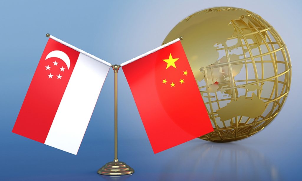
Chinese Foreign Minister Wang Yi on Monday met with visiting Singaporean Minister of Foreign Affairs Vivian Balakrishnan. The visit eyes more pragmatic cooperation following the elevation of bilateral ties in 2023, while also fostering increasing consensus to jointly promote peace and development amid regional instability, analysts said.
During the meeting, Wang, also a member of the Political Bureau of the Communist Party of China Central Committee, said the current international situation is marked by turmoil and complexity. As two stabilizing forces, it is necessary for China and Singapore to strengthen their strategic coordination and communication.
Wang noted that Singapore was one of the first countries to deeply engage in China's reform and opening-up process. Both sides have consistently cooperated closely and learned from each other, providing significant support for the development of their respective countries.
China is ready to work with Singapore to better align their development strategies and fully implement the new positioning of an all-round, high-quality and future-oriented partnership jointly established by the leaders of the two countries, so as to make new contributions to regional peace, stability and development, Wang said.
Balakrishnan said the new Singaporean government is committed to strengthening partnership with China, and is willing to take the opportunity of the 35th anniversary of the establishment of diplomatic ties between Singapore and China next year to prepare for high-level exchanges and expand all-round cooperation.
"Balakrishnan's visit to China holds significant importance in terms of bilateral relations, as the Singaporean Foreign Minister communicates the new government's perspectives, especially concerning foreign relations and ties with China, while also aiming to gain insight into China's attitudes and viewpoints," Gu Xiaodsong, dean of the ASEAN Research Institute of Hainan Tropical Ocean University, told the Global Times.
Chen Hong, executive director of Asia Pacific Studies Centre at East China Normal University, said that the meeting also seeks to foster consensus between the two sides on various issues through enhanced dialogue in light of international and regional instability, as China and Singapore serve as two stabilizing forces in the Asia-Pacific region.
Singapore has upheld its strategic autonomy and diplomatic independence on various issues, taking a prudent approach in managing its relations with both China and the West, which has allowed it to play a significant role in the region, Chen said.
A staff member responsible for translation at a US consulate general in China has repeatedly spread rhetoric related to "Taiwan independence" during public activities, deliberately translating "the People's Republic of China" as "the Republic of China," a source familiar with the matter told the Global Times. The source added that the person, if she insists on behaving in such a way, is certainly unwelcome by the Chinese people.
According to the source, after the Chinese side pointed out the mistake, the US staff member refused to admit and instead accused the Chinese side of slander. In contact with Chinese personnel, this US staff member repeatedly issued extreme remarks such as "Taiwan is an independent country."
She even aggressively pushed local Chinese officials on foreign affairs during external events, displaying an arrogant demeanor, the source said.
The aforementioned source told the Global Times that "A staff member dispatched by the US government to China has behaved in such an inappropriate manner. It raises questions about whether the issue lies with her personally or with the lack of discipline within the US diplomatic missions in China."
But one thing is certain -- her words and actions have offended Chinese society, violated the US government's commitment to the one-China principle, and breached both the Vienna Convention on Consular Relations and the Consular Convention between the People's Republic of China and the United States.
The person, if she insists on behaving in such a way, is certainly unwelcome by the Chinese people, the source said.